 Movie
Movie Controller


+ Open data

- Basic information
| Entry | Database: PDB / ID: 4eba | ||||||
|---|---|---|---|---|---|---|---|
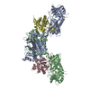
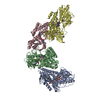
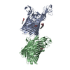
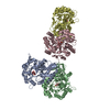
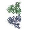
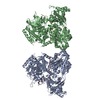
| Title | Crystal structure of the Rna14-Rna15 complex | ||||||
 Components Components |
| ||||||
 Keywords Keywords | STRUCTURAL PROTEIN/RNA BINDING PROTEIN / HAT domain / HEAT repeat / monkeytail / Clp1 / Pcf11 / STRUCTURAL PROTEIN-RNA BINDING PROTEIN complex | ||||||
| Function / homology |  Function and homology information Function and homology informationmRNA cleavage and polyadenylation specificity factor complex / mRNA 3'-end processing / mRNA binding / nucleus / cytoplasm Similarity search - Function | ||||||
| Biological species |  Kluyveromyces lactis (yeast) Kluyveromyces lactis (yeast) | ||||||
| Method |  X-RAY DIFFRACTION / SYNCHROTRON / MOLECULAR REPLACEMENT / Resolution: 3.3 Å X-RAY DIFFRACTION / SYNCHROTRON / MOLECULAR REPLACEMENT / Resolution: 3.3 Å | ||||||
 Authors Authors | Paulson, A.R. / Tong, L. | ||||||
 Citation Citation | Journal: Rna / Year: 2012 Title: Crystal structure of the Rna14-Rna15 complex. Authors: Paulson, A.R. / Tong, L. | ||||||
| History |
|
- Structure visualization
| Structure viewer | Molecule: MolmilJmol/JSmol |
|---|
- Downloads & links
-Download
| PDBx/mmCIF format | 4eba.cif.gz | 733.1 KB | Display | PDBx/mmCIF format |
|---|---|---|---|---|
| PDB format | pdb4eba.ent.gz | 603.5 KB | Display | PDB format |
| PDBx/mmJSON format | 4eba.json.gz | Tree view | PDBx/mmJSON format | |
| Others |  Other downloads Other downloads |
-Validation report
| Arichive directory | https://data.pdbj.org/pub/pdb/validation_reports/eb/4ebaftp://data.pdbj.org/pub/pdb/validation_reports/eb/4eba | HTTPS FTP |
|---|
-Related structure data
| Related structure data |  4e6hC  4e85SC C: citing same article ( S: Starting model for refinement |
|---|---|
| Similar structure data |


-Links
 PDBj
PDBj
- Assembly
| Deposited unit | 
| ||||||||
|---|---|---|---|---|---|---|---|---|---|
| 1 | 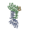
| ||||||||
| 2 | 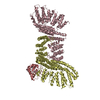
| ||||||||
| 3 | 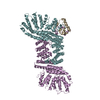
| ||||||||
| Unit cell |
| ||||||||
| Details | The trimers are formed by two Rna14 and one Rna15. |
-Components
| #1: Protein | Mass: 76161.523 Da / Num. of mol.: 6 / Fragment: UNP residues 18-661 Source method: isolated from a genetically manipulated source Source: (gene. exp.) Kluyveromyces lactis (yeast)Strain: ATCC 8585 / CBS 2359 / DSM 70799 / NBRC 1267 / NRRL Y-1140 / WM37 Gene: RNA14, KLLA0F26290g / Production host:  #2: Protein | Mass: 19549.305 Da / Num. of mol.: 3 / Fragment: UNP residues 105-258 Source method: isolated from a genetically manipulated source Source: (gene. exp.) Kluyveromyces lactis (yeast)Strain: ATCC 8585 / CBS 2359 / DSM 70799 / NBRC 1267 / NRRL Y-1140 / WM37 Gene: KLLA0F09383g / Production host: |
|---|
-Experimental details
-Experiment
| Experiment | Method: X-RAY DIFFRACTION / Number of used crystals: 1 |
|---|
- Sample preparation
| Crystal | Density Matthews: 2.61 Å3/Da / Density % sol: 52.87 % |
|---|---|
| Crystal grow | Temperature: 293 K / Method: vapor diffusion, hanging drop / pH: 7 Details: 50 mM HEPES 7.0, 1% tryptone, 13% PEG 3350, VAPOR DIFFUSION, HANGING DROP, temperature 293K |
-Data collection
| Diffraction | Mean temperature: 100 K |
|---|---|
| Diffraction source | Source: SYNCHROTRON / Site: NSLS  / Beamline: X29A / Wavelength: 1.075 / Wavelength: 1.075 Å / Beamline: X29A / Wavelength: 1.075 / Wavelength: 1.075 Å |
| Detector | Type: ADSC QUANTUM 315 / Detector: CCD / Date: Nov 9, 2011 / Details: MIRRORS |
| Radiation | Monochromator: SI(111) / Protocol: SINGLE WAVELENGTH / Monochromatic (M) / Laue (L): M / Scattering type: x-ray |
| Radiation wavelength | Wavelength: 1.075 Å / Relative weight: 1 |
| Reflection | Resolution: 3.3→50 Å / Num. all: 79000 / Num. obs: 71640 / % possible obs: 90.9 % / Observed criterion σ(F): 0 / Observed criterion σ(I): -3 / Redundancy: 1.7 % / Rmerge(I) obs: 0.055 / Net I/σ(I): 10.9049 |
| Reflection shell | Resolution: 3.3→3.42 Å / Redundancy: 1.5 % / Rmerge(I) obs: 0.442 / Mean I/σ(I) obs: 1.242 / % possible all: 71.2 |
- Processing
| Software |
| ||||||||||||||||||||||||||||||||||||||||||||||||||||||||||||
|---|---|---|---|---|---|---|---|---|---|---|---|---|---|---|---|---|---|---|---|---|---|---|---|---|---|---|---|---|---|---|---|---|---|---|---|---|---|---|---|---|---|---|---|---|---|---|---|---|---|---|---|---|---|---|---|---|---|---|---|---|---|
| Refinement | Method to determine structure: MOLECULAR REPLACEMENT Starting model: PDB ENTRY 4E85 Resolution: 3.3→47.79 Å / Rfactor Rfree error: 0.005 / Data cutoff high absF: 371325.06 / Data cutoff low absF: 0 / Isotropic thermal model: RESTRAINED / Cross valid method: THROUGHOUT / σ(F): 0 / Details: BULK SOLVENT MODEL USED
| ||||||||||||||||||||||||||||||||||||||||||||||||||||||||||||
| Solvent computation | Solvent model: FLAT MODEL / Bsol: 57.2895 Å2 / ksol: 0.3 e/Å3 | ||||||||||||||||||||||||||||||||||||||||||||||||||||||||||||
| Displacement parameters | Biso mean: 100.2 Å2
| ||||||||||||||||||||||||||||||||||||||||||||||||||||||||||||
| Refine analyze |
| ||||||||||||||||||||||||||||||||||||||||||||||||||||||||||||
| Refinement step | Cycle: LAST / Resolution: 3.3→47.79 Å
| ||||||||||||||||||||||||||||||||||||||||||||||||||||||||||||
| Refine LS restraints |
| ||||||||||||||||||||||||||||||||||||||||||||||||||||||||||||
| Refine LS restraints NCS | NCS model details: CONSTR | ||||||||||||||||||||||||||||||||||||||||||||||||||||||||||||
| LS refinement shell | Resolution: 3.3→3.42 Å / Rfactor Rfree error: 0.027 / Total num. of bins used: 10
| ||||||||||||||||||||||||||||||||||||||||||||||||||||||||||||
| Xplor file | Serial no: 1 / Param file: protein_rep.param / Topol file: protein.top |
