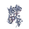
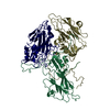
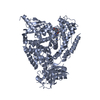
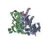
[English] 日本語
Yorodumi
- PDB-3rnm: The crystal structure of the subunit binding of human dihydrolipo... -

+
Open data


ID or keywords:

Loading...

-
Basic information

Entry
Database: PDB / ID: 3rnm
TitleThe crystal structure of the subunit binding of human dihydrolipoamide transacylase (E2b) bound to human dihydrolipoamide dehydrogenase (E3)
Components
  • Dihydrolipoyl dehydrogenase, mitochondrialDihydrolipoamide dehydrogenase
  • Lipoamide acyltransferase component of branched-chain alpha-keto acid dehydrogenase complex, mitochondrial
KeywordsOXIDOREDUCTASE/PROTEIN BINDING / protein-protein interaction / redox protein / mitochondrion / OXIDOREDUCTASE-PROTEIN BINDING complex
Function / homology
Function and homology information


dihydrolipoyllysine-residue (2-methylpropanoyl)transferase / dihydrolipoyllysine-residue (2-methylpropanoyl)transferase activity / acetyltransferase complex / acrosomal matrix / Glycine degradation / : / oxoglutarate dehydrogenase complex / dihydrolipoyl dehydrogenase / dihydrolipoyl dehydrogenase activity / : ...dihydrolipoyllysine-residue (2-methylpropanoyl)transferase / dihydrolipoyllysine-residue (2-methylpropanoyl)transferase activity / acetyltransferase complex / acrosomal matrix / Glycine degradation / : / oxoglutarate dehydrogenase complex / dihydrolipoyl dehydrogenase / dihydrolipoyl dehydrogenase activity / : / Lysine catabolism / acetyl-CoA biosynthetic process from pyruvate / lipoic acid binding / pyruvate dehydrogenase complex / branched-chain amino acid catabolic process / : / Citric acid cycle (TCA cycle) / Branched-chain amino acid catabolism / Pyruvate metabolism / Glyoxylate metabolism and glycine degradation / Regulation of pyruvate dehydrogenase (PDH) complex / motile cilium / sperm capacitation / Signaling by Retinoic Acid / mitochondrial nucleoid / mitochondrial electron transport, NADH to ubiquinone / acetyltransferase activity / RHOH GTPase cycle / gastrulation / regulation of membrane potential / microtubule cytoskeleton / flavin adenine dinucleotide binding / mitochondrial matrix / ubiquitin protein ligase binding / mitochondrion / proteolysis / nucleus / cytosol / cytoplasm
Similarity search - Function
E3-binding domain / Dihydrolipoamide Transferase / Dihydrolipoamide dehydrogenase / Peripheral subunit-binding domain / e3 binding domain / Peripheral subunit-binding (PSBD) domain profile. / E3-binding domain superfamily / 2-oxo acid dehydrogenase, lipoyl-binding site / 2-oxo acid dehydrogenases acyltransferase component lipoyl binding site. / 2-oxoacid dehydrogenase acyltransferase, catalytic domain ...E3-binding domain / Dihydrolipoamide Transferase / Dihydrolipoamide dehydrogenase / Peripheral subunit-binding domain / e3 binding domain / Peripheral subunit-binding (PSBD) domain profile. / E3-binding domain superfamily / 2-oxo acid dehydrogenase, lipoyl-binding site / 2-oxo acid dehydrogenases acyltransferase component lipoyl binding site. / 2-oxoacid dehydrogenase acyltransferase, catalytic domain / 2-oxoacid dehydrogenases acyltransferase (catalytic domain) / Pyridine nucleotide-disulphide oxidoreductase, class I / FAD/NAD-linked reductase, C-terminal dimerisation domain / Pyridine nucleotide-disulphide oxidoreductase, class I, active site / Pyridine nucleotide-disulphide oxidoreductases class-I active site. / Biotin-requiring enzyme / Pyridine nucleotide-disulphide oxidoreductase, dimerisation domain / Pyridine nucleotide-disulphide oxidoreductase, dimerisation domain / Biotinyl/lipoyl domain profile. / Biotin/lipoyl attachment / Single hybrid motif / FAD/NAD-linked reductase, dimerisation domain superfamily / Chloramphenicol acetyltransferase-like domain superfamily / FAD/NAD(P)-binding domain / Pyridine nucleotide-disulphide oxidoreductase / Enolase-like; domain 1 / FAD/NAD(P)-binding domain / FAD/NAD(P)-binding domain / 3-Layer(bba) Sandwich / FAD/NAD(P)-binding domain superfamily / Few Secondary Structures / Irregular / 2-Layer Sandwich / Alpha Beta
Similarity search - Domain/homology
BETA-MERCAPTOETHANOL / FLAVIN-ADENINE DINUCLEOTIDE / Dihydrolipoyl dehydrogenase, mitochondrial / Lipoamide acyltransferase component of branched-chain alpha-keto acid dehydrogenase complex, mitochondrial
Similarity search - Component
Biological speciesHomo sapiens (human)
MethodX-RAY DIFFRACTION / SYNCHROTRON / FOURIER SYNTHESIS / Resolution: 2.4 Å
AuthorsBrautigam, C.A. / Wynn, R.M. / Chuang, J.C. / Young, B.B. / Chuang, D.T.
CitationJournal: J.Biol.Chem. / Year: 2011
Title: Structural and Thermodynamic Basis for Weak Interactions between Dihydrolipoamide Dehydrogenase and Subunit-binding Domain of the Branched-chain {alpha}-Ketoacid Dehydrogenase Complex.
Authors: Brautigam, C.A. / Wynn, R.M. / Chuang, J.L. / Naik, M.T. / Young, B.B. / Huang, T.H. / Chuang, D.T.
History
DepositionApr 22, 2011Deposition site: RCSB / Processing site: RCSB
Revision 1.0May 4, 2011Provider: repository / Type: Initial release
Revision 1.1Jul 13, 2011Group: Version format compliance
Revision 1.2Jul 20, 2011Group: Database references

-
Structure visualization

Structure viewerMolecule:
MolmilJmol/JSmol

Downloads & links

-
Assembly

Deposited unit
A: Dihydrolipoyl dehydrogenase, mitochondrial
B: Dihydrolipoyl dehydrogenase, mitochondrial
C: Dihydrolipoyl dehydrogenase, mitochondrial
D: Dihydrolipoyl dehydrogenase, mitochondrial
E: Lipoamide acyltransferase component of branched-chain alpha-keto acid dehydrogenase complex, mitochondrial
F: Lipoamide acyltransferase component of branched-chain alpha-keto acid dehydrogenase complex, mitochondrial
hetero molecules


Theoretical massNumber of molelcules
Total (without water)228,07718
Polymers223,7946
Non-polymers4,28412
Water6,251347
1
A: Dihydrolipoyl dehydrogenase, mitochondrial
B: Dihydrolipoyl dehydrogenase, mitochondrial
E: Lipoamide acyltransferase component of branched-chain alpha-keto acid dehydrogenase complex, mitochondrial
hetero molecules


Theoretical massNumber of molelcules
Total (without water)114,0399
Polymers111,8973
Non-polymers2,1426
Water543
TypeNameSymmetry operationNumber
identity operation1_555x,y,z1
2
C: Dihydrolipoyl dehydrogenase, mitochondrial
D: Dihydrolipoyl dehydrogenase, mitochondrial
F: Lipoamide acyltransferase component of branched-chain alpha-keto acid dehydrogenase complex, mitochondrial
hetero molecules


Theoretical massNumber of molelcules
Total (without water)114,0399
Polymers111,8973
Non-polymers2,1426
Water543
TypeNameSymmetry operationNumber
identity operation1_555x,y,z1
Unit cell
Length a, b, c (Å)84.136, 112.308, 122.545
Angle α, β, γ (deg.)90.00, 91.40, 90.00
Int Tables number4
Space group name H-MP1211
DetailsChains A, B, and E / Chains C, D, and F

-
Components

-
Protein , 2 types, 6 molecules ABCDEF

#1: Protein
Dihydrolipoyl dehydrogenase, mitochondrial / Dihydrolipoamide dehydrogenase / Dihydrolipoamide dehydrogenase / Glycine cleavage system L protein


Mass: 52652.137 Da / Num. of mol.: 4 / Fragment: Residues 36-509
Source method: isolated from a genetically manipulated source
Source: (gene. exp.) Homo sapiens (human) / Gene: DLD, GCSL, LAD, PHE3 / Production host: Escherichia coli (E. coli) / References: UniProt: P09622, dihydrolipoyl dehydrogenase
#2: Protein Lipoamide acyltransferase component of branched-chain alpha-keto acid dehydrogenase complex, mitochondrial / Branched-chain alpha-keto acid dehydrogenase complex component E2 / BCKAD-E2 / BCKADE2 / ...Branched-chain alpha-keto acid dehydrogenase complex component E2 / BCKAD-E2 / BCKADE2 / Dihydrolipoamide acetyltransferase component of branched-chain alpha-keto acid dehydrogenase complex / Dihydrolipoamide branched chain transacylase / Dihydrolipoyllysine-residue (2-methylpropanoyl)transferase


Mass: 6592.526 Da / Num. of mol.: 2 / Fragment: subunit-binding domain, residues 165-213 / Mutation: R118N
Source method: isolated from a genetically manipulated source
Source: (gene. exp.) Homo sapiens (human) / Gene: DBT, BCATE2 / Production host: Escherichia coli (E. coli)
References: UniProt: P11182, dihydrolipoyllysine-residue (2-methylpropanoyl)transferase

-
Non-polymers , 4 types, 359 molecules

#3: Chemical
ChemComp-BME / BETA-MERCAPTOETHANOL / 2-Mercaptoethanol


Mass: 78.133 Da / Num. of mol.: 4 / Source method: obtained synthetically / Formula: C2H6OS
#4: Chemical
ChemComp-FAD / FLAVIN-ADENINE DINUCLEOTIDE / Flavin adenine dinucleotide


Mass: 785.550 Da / Num. of mol.: 4 / Source method: obtained synthetically / Formula: C27H33N9O15P2 / Comment: FAD*YM
#5: Chemical
ChemComp-NHE / 2-[N-CYCLOHEXYLAMINO]ETHANE SULFONIC ACID / N-CYCLOHEXYLTAURINE / CHES / CHES (buffer)


Mass: 207.290 Da / Num. of mol.: 4 / Source method: obtained synthetically / Formula: C8H17NO3S / Comment: pH buffer*YM
#6: Water ChemComp-HOH / water / Water


Mass: 18.015 Da / Num. of mol.: 347 / Source method: isolated from a natural source / Formula: H2O

-
Experimental details

-
Experiment

ExperimentMethod: X-RAY DIFFRACTION / Number of used crystals: 1

-
Sample preparation

CrystalDensity Matthews: 2.58 Å3/Da / Density % sol: 52.36 %
Crystal growTemperature: 293 K / Method: vapor diffusion, hanging drop / pH: 9.1
Details: 18% PEG3350 0.1 M CHES , pH 9.1, VAPOR DIFFUSION, HANGING DROP, temperature 293K

-
Data collection

DiffractionMean temperature: 100 K
Diffraction sourceSource: SYNCHROTRON / Site: APS / Beamline: 19-ID / Wavelength: 0.97918 Å
DetectorType: ADSC QUANTUM 315r / Detector: CCD / Date: Apr 19, 2010
RadiationMonochromator: Si 111 / Protocol: SINGLE WAVELENGTH / Monochromatic (M) / Laue (L): M / Scattering type: x-ray
Radiation wavelengthWavelength: 0.97918 Å / Relative weight: 1
ReflectionResolution: 2.4→50 Å / Num. all: 88688 / Num. obs: 88688 / % possible obs: 100 % / Observed criterion σ(I): -3
Reflection shellResolution: 2.4→2.44 Å / % possible all: 99.9

-
Processing

Software
NameVersionClassification
SBC-Collectdata collection
PHENIXmodel building
PHENIX(phenix.refine: 1.7_650)refinement
HKL-3000data reduction
HKL-3000data scaling
PHENIXphasing
RefinementMethod to determine structure: FOURIER SYNTHESIS / Resolution: 2.4→44.872 Å / SU ML: 0.39 / σ(F): 0 / Phase error: 25.97 / Stereochemistry target values: ML
RfactorNum. reflection% reflectionSelection details
Rfree0.2395 4446 5.02 %random
Rwork0.1814 ---
obs0.1843 88635 99.28 %-
all-88635 --
Solvent computationShrinkage radii: 0.95 Å / VDW probe radii: 1.2 Å / Solvent model: FLAT BULK SOLVENT MODEL / Bsol: 29.59 Å2 / ksol: 0.309 e/Å3
Displacement parameters
Baniso -1Baniso -2Baniso -3
1--6.8722 Å20 Å2-2.7522 Å2
2--8.26 Å20 Å2
3----1.3878 Å2
Refinement stepCycle: LAST / Resolution: 2.4→44.872 Å
ProteinNucleic acidLigandSolventTotal
Num. atoms14720 0 280 347 15347
Refine LS restraints
Refine-IDTypeDev idealNumber
X-RAY DIFFRACTIONf_bond_d0.00815262
X-RAY DIFFRACTIONf_angle_d1.08220639
X-RAY DIFFRACTIONf_dihedral_angle_d13.775615
X-RAY DIFFRACTIONf_chiral_restr0.0682378
X-RAY DIFFRACTIONf_plane_restr0.0042622
LS refinement shell
Resolution (Å)Rfactor RfreeNum. reflection RfreeRfactor RworkNum. reflection RworkRefine-ID% reflection obs (%)
2.4-2.42550.38871230.30162222X-RAY DIFFRACTION80
2.4255-2.4540.35061330.26992816X-RAY DIFFRACTION100
2.454-2.48390.29961400.25752863X-RAY DIFFRACTION100
2.4839-2.51540.32231500.25542783X-RAY DIFFRACTION100
2.5154-2.54850.30911560.23742857X-RAY DIFFRACTION100
2.5485-2.58340.3221420.23512768X-RAY DIFFRACTION100
2.5834-2.62030.29121490.23662807X-RAY DIFFRACTION100
2.6203-2.65940.34321460.22522819X-RAY DIFFRACTION100
2.6594-2.70090.31421310.21182838X-RAY DIFFRACTION100
2.7009-2.74520.27051540.20882811X-RAY DIFFRACTION100
2.7452-2.79250.30631610.21222815X-RAY DIFFRACTION100
2.7925-2.84330.26451620.2142816X-RAY DIFFRACTION100
2.8433-2.8980.29181590.20642801X-RAY DIFFRACTION100
2.898-2.95710.32161420.20452808X-RAY DIFFRACTION100
2.9571-3.02140.2861420.20852859X-RAY DIFFRACTION100
3.0214-3.09170.25691440.19252776X-RAY DIFFRACTION100
3.0917-3.1690.24841420.20022823X-RAY DIFFRACTION100
3.169-3.25460.2551440.20362878X-RAY DIFFRACTION100
3.2546-3.35040.28041440.19542821X-RAY DIFFRACTION100
3.3504-3.45850.24451310.20362820X-RAY DIFFRACTION100
3.4585-3.58210.25841670.19772818X-RAY DIFFRACTION100
3.5821-3.72540.27241540.19622819X-RAY DIFFRACTION100
3.7254-3.89490.25231500.17912838X-RAY DIFFRACTION100
3.8949-4.10010.20661270.15362835X-RAY DIFFRACTION100
4.1001-4.35680.17281480.13372839X-RAY DIFFRACTION100
4.3568-4.69280.18541730.12212840X-RAY DIFFRACTION100
4.6928-5.16450.16611450.13082823X-RAY DIFFRACTION100
5.1645-5.91030.20031410.16222864X-RAY DIFFRACTION100
5.9103-7.44090.20341700.16672841X-RAY DIFFRACTION100
7.4409-44.87980.18831760.15142871X-RAY DIFFRACTION99
Refinement TLS params.

Method: refined / Refine-ID: X-RAY DIFFRACTION

IDL112)L122)L132)L222)L232)L332)S11 (Å °)S12 (Å °)S13 (Å °)S21 (Å °)S22 (Å °)S23 (Å °)S31 (Å °)S32 (Å °)S33 (Å °)T112)T122)T132)T222)T232)T332)Origin x (Å)Origin y (Å)Origin z (Å)
10.29440.117-0.06630.28630.07390.26970.0078-0.03310.0182-0.0078-0.08460.053-0.0589-0.01120.0520.06210.0097-0.00270.0642-0.00850.0459-18.03472.897631.3639
20.04270.03150.01110.1049-0.1170.2317-0.00640.0214-0.0134-0.0078-0.1384-0.1758-0.02750.28140.1340.0864-0.02140.01610.31970.15290.18416.61137.489624.4501
30.3977-0.17130.1980.2118-0.09550.31020.04120.04210.072-0.01440.0033-0.05620.06160.0204-0.01960.1020.0387-0.00390.07760.01980.089-21.115-50.412439.0791
40.4045-0.08020.31980.3556-0.03430.25430.056-0.061-0.0049-0.0679-0.05360.0860.0252-0.1038-0.00450.09630.0256-0.0330.14250.02990.0812-55.7099-49.306230.3869
50.74260.4267-0.26350.64840.46711.04150.05740.00530.06950.0850.0606-0.23910.08070.0173-0.11470.1875-0.00890.01060.166-0.00930.2412-4.073641.506831.5875
60.13740.0051-0.00250.0440.08820.17780.00010.06080.0715-0.4072-0.0987-0.0671-0.1931-0.03560.09830.6940.0354-0.05330.52710.02390.7117-36.5924-83.335630.6687
Refinement TLS group
IDRefine-IDRefine TLS-IDSelection details
1X-RAY DIFFRACTION1Chain A
2X-RAY DIFFRACTION2Chain B
3X-RAY DIFFRACTION3Chain C
4X-RAY DIFFRACTION4Chain D
5X-RAY DIFFRACTION5Chain E
6X-RAY DIFFRACTION6Chain F

+
About Yorodumi

-
News

-
Feb 9, 2022. New format data for meta-information of EMDB entries

New format data for meta-information of EMDB entries

  • Version 3 of the EMDB header file is now the official format.
  • The previous official version 1.9 will be removed from the archive.

Related info.:EMDB header

External links:wwPDB to switch to version 3 of the EMDB data model

-
Aug 12, 2020. Covid-19 info

Covid-19 info

URL: https://pdbj.org/emnavi/covid19.php

New page: Covid-19 featured information page in EM Navigator.

Related info.:Covid-19 info / Mar 5, 2020. Novel coronavirus structure data

+
Mar 5, 2020. Novel coronavirus structure data

Novel coronavirus structure data

Related info.:Yorodumi Speices / Aug 12, 2020. Covid-19 info

External links:COVID-19 featured content - PDBj / Molecule of the Month (242):Coronavirus Proteases

+
Jan 31, 2019. EMDB accession codes are about to change! (news from PDBe EMDB page)

EMDB accession codes are about to change! (news from PDBe EMDB page)

  • The allocation of 4 digits for EMDB accession codes will soon come to an end. Whilst these codes will remain in use, new EMDB accession codes will include an additional digit and will expand incrementally as the available range of codes is exhausted. The current 4-digit format prefixed with “EMD-” (i.e. EMD-XXXX) will advance to a 5-digit format (i.e. EMD-XXXXX), and so on. It is currently estimated that the 4-digit codes will be depleted around Spring 2019, at which point the 5-digit format will come into force.
  • The EM Navigator/Yorodumi systems omit the EMD- prefix.

Related info.:Q: What is EMD? / ID/Accession-code notation in Yorodumi/EM Navigator

External links:EMDB Accession Codes are Changing Soon! / Contact to PDBj

+
Jul 12, 2017. Major update of PDB

Major update of PDB

  • wwPDB released updated PDB data conforming to the new PDBx/mmCIF dictionary.
  • This is a major update changing the version number from 4 to 5, and with Remediation, in which all the entries are updated.
  • In this update, many items about electron microscopy experimental information are reorganized (e.g. em_software).
  • Now, EM Navigator and Yorodumi are based on the updated data.

External links:wwPDB Remediation / Enriched Model Files Conforming to OneDep Data Standards Now Available in the PDB FTP Archive

-
Yorodumi

Thousand views of thousand structures

  • Yorodumi is a browser for structure data from EMDB, PDB, SASBDB, etc.
  • This page is also the successor to EM Navigator detail page, and also detail information page/front-end page for Omokage search.
  • The word "yorodu" (or yorozu) is an old Japanese word meaning "ten thousand". "mi" (miru) is to see.

Related info.:EMDB / PDB / SASBDB / Comparison of 3 databanks / Yorodumi Search / Aug 31, 2016. New EM Navigator & Yorodumi / Yorodumi Papers / Jmol/JSmol / Function and homology information / Changes in new EM Navigator and Yorodumi

Read more