Movie
Movie Controller

[English] 日本語
 Yorodumi
- EMDB-1646: Structure of the Human Dicer-TRBP Complex by Electron Microscopy -
+ Open data

- Basic information
| Entry | Database: EMDB / ID: EMD-1646 | |||||||||
|---|---|---|---|---|---|---|---|---|---|---|
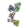
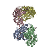
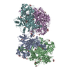
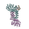
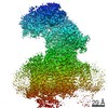
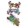
| Title | Structure of the Human Dicer-TRBP Complex by Electron Microscopy | |||||||||
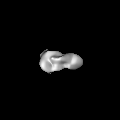
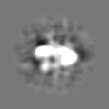
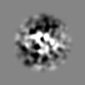
 Map data Map data | This is the 3D volume of human Dicer-TRBP complex | |||||||||
 Sample Sample |
| |||||||||
 Keywords Keywords | Dicer Enzyme / Ribonuclease III / MicroRNA Processing / Single Particle Electron Microscopy | |||||||||
| Biological species |  Homo sapiens (human) Homo sapiens (human) | |||||||||
| Method | single particle reconstruction / negative staining / Resolution: 20.0 Å | |||||||||
 Authors Authors | Lau PW / Potter CS / Carragher B / MacRae IJ | |||||||||
 Citation Citation | Journal: Structure / Year: 2009 Title: Structure of the human Dicer-TRBP complex by electron microscopy. Authors: Pick-Wei Lau / Clinton S Potter / Bridget Carragher / Ian J MacRae /  Abstract: Dicer is a specialized ribonuclease that initiates RNA interference (RNAi) by cleaving double-stranded RNA (dsRNA) into small RNA fragments about 22 nucleotides long. Here, we present the three- ...Dicer is a specialized ribonuclease that initiates RNA interference (RNAi) by cleaving double-stranded RNA (dsRNA) into small RNA fragments about 22 nucleotides long. Here, we present the three-dimensional structure of human Dicer bound to the protein TRBP at approximately 20 A resolution determined by negative-stain electron microscopy (EM) and single-particle analysis. Our analysis reveals that the Dicer-TRBP complex is an L-shaped molecule with a long edge of 150 A and a 100 A extension on one end. A surface trench runs the length of the long edge of the molecule, defining a putative dsRNA-binding site. Docking the crystal structure of Giardia Dicer, which represents the nuclease core of human Dicer, into the EM map suggests two possible overall molecular architectures for human Dicer. These results offer insights into the structure of Dicer proteins found in multicellular organisms and provide a conceptual framework for understanding the initiation of RNAi. | |||||||||
| History |
|
- Structure visualization
| Movie |
 Movie viewer Movie viewer |
|---|---|
| Structure viewer | EM map: SurfViewMolmilJmol/JSmol |
| Supplemental images |

- Downloads & links
-EMDB archive
| Map data | emd_1646.map.gz | 4.5 MB | EMDB map data format | |
|---|---|---|---|---|
| Header (meta data) | emd-1646-v30.xmlemd-1646.xml | 9.8 KB 9.8 KB | Display Display | EMDB header |
| Images | 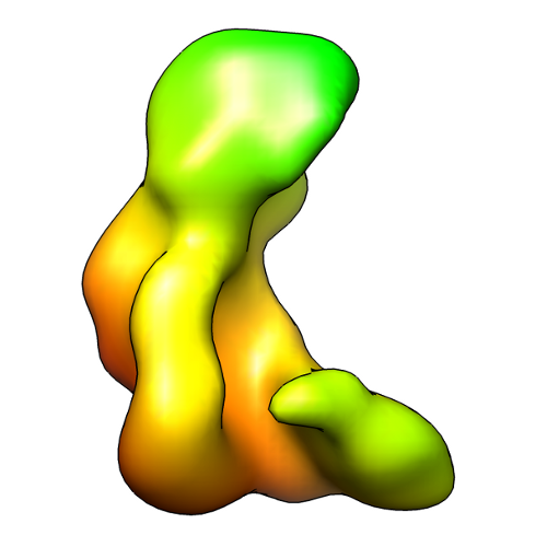 EMD-1646_image.png EMD-1646_image.png | 104 KB | ||
| Archive directory |  http://ftp.pdbj.org/pub/emdb/structures/EMD-1646ftp://ftp.pdbj.org/pub/emdb/structures/EMD-1646 http://ftp.pdbj.org/pub/emdb/structures/EMD-1646ftp://ftp.pdbj.org/pub/emdb/structures/EMD-1646 | HTTPS FTP |
-Validation report
| Summary document | emd_1646_validation.pdf.gz | 186.4 KB | Display | EMDB validaton report |
|---|---|---|---|---|
| Full document | emd_1646_full_validation.pdf.gz | 185.5 KB | Display | |
| Data in XML | emd_1646_validation.xml.gz | 5.5 KB | Display | |
| Arichive directory | https://ftp.pdbj.org/pub/emdb/validation_reports/EMD-1646ftp://ftp.pdbj.org/pub/emdb/validation_reports/EMD-1646 | HTTPS FTP |
-Related structure data
| Similar structure data |
|---|
-Links
| EMDB pages | EMDB (EBI/PDBe) / EMDataResource |
|---|
-Map
| File | Download / File: emd_1646.map.gz / Format: CCP4 / Size: 6.4 MB / Type: IMAGE STORED AS FLOATING POINT NUMBER (4 BYTES) | ||||||||||||||||||||||||||||||||||||||||||||||||||||||||||||||||||||
|---|---|---|---|---|---|---|---|---|---|---|---|---|---|---|---|---|---|---|---|---|---|---|---|---|---|---|---|---|---|---|---|---|---|---|---|---|---|---|---|---|---|---|---|---|---|---|---|---|---|---|---|---|---|---|---|---|---|---|---|---|---|---|---|---|---|---|---|---|---|
| Annotation | This is the 3D volume of human Dicer-TRBP complex | ||||||||||||||||||||||||||||||||||||||||||||||||||||||||||||||||||||
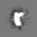
| Projections & slices | Image control
Images are generated by Spider. | ||||||||||||||||||||||||||||||||||||||||||||||||||||||||||||||||||||
| Voxel size | X=Y=Z: 3.093 Å | ||||||||||||||||||||||||||||||||||||||||||||||||||||||||||||||||||||
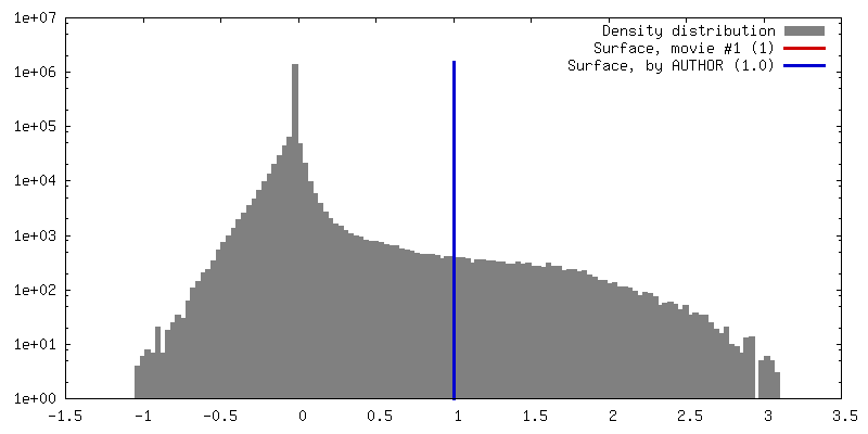
| Density |
| ||||||||||||||||||||||||||||||||||||||||||||||||||||||||||||||||||||
| Symmetry | Space group: 1 | ||||||||||||||||||||||||||||||||||||||||||||||||||||||||||||||||||||
| Details | EMDB XML:
CCP4 map header:
| ||||||||||||||||||||||||||||||||||||||||||||||||||||||||||||||||||||
 Z (Sec.)
 Y (Row.)
 X (Col.)


-Supplemental data
- Sample components
-Entire : Dicer-TRBP complex
| Entire | Name: Dicer-TRBP complex |
|---|---|
| Components |
|
-Supramolecule #1000: Dicer-TRBP complex
| Supramolecule | Name: Dicer-TRBP complex / type: sample / ID: 1000 / Details: The sample was mostly monodisperse Oligomeric state: One human Dicer binds to one TRBP molecule Number unique components: 2 |
|---|---|
| Molecular weight | Theoretical: 250 KDa |
-Macromolecule #1: Human Dicer
| Macromolecule | Name: Human Dicer / type: protein_or_peptide / ID: 1 / Name.synonym: hDicer / Number of copies: 1 / Oligomeric state: Monomer / Recombinant expression: Yes |
|---|---|
| Source (natural) | Organism: Homo sapiens (human) / synonym: Human |
| Molecular weight | Theoretical: 220 KDa |
| Recombinant expression | Organism:   Spodoptera frugiperda (fall armyworm) / Recombinant plasmid: Baculovirus Spodoptera frugiperda (fall armyworm) / Recombinant plasmid: Baculovirus |
-Macromolecule #2: TAR RNA binding protein
| Macromolecule | Name: TAR RNA binding protein / type: protein_or_peptide / ID: 2 / Name.synonym: TRBP / Number of copies: 1 / Oligomeric state: Monomer / Recombinant expression: Yes |
|---|---|
| Source (natural) | Organism: Homo sapiens (human) / synonym: Human |
| Molecular weight | Theoretical: 30 KDa |
| Recombinant expression | Organism: Spodoptera frugiperda (fall armyworm) / Recombinant plasmid: Baculovirus |
-Experimental details
-Structure determination
| Method | negative staining |
|---|---|
 Processing Processing | single particle reconstruction |
| Aggregation state | particle |
-Sample preparation
| Buffer | pH: 7.4 / Details: 150mM KCl, 20mM HEPES |
|---|---|
| Staining | Type: NEGATIVE Details: Grids with adsorbed protein floated on 2% w/v uranyl acetate for approximately 20 seconds. |
| Vitrification | Cryogen name: NONE / Instrument: OTHER |
- Electron microscopy
| Microscope | FEI TECNAI F20 |
|---|---|
| Date | May 31, 2009 |
| Image recording | Category: CCD / Film or detector model: TVIPS TEMCAM-F415 (4k x 4k) / Number real images: 947 / Average electron dose: 20 e/Å2 |
| Electron beam | Acceleration voltage: 120 kV / Electron source:  FIELD EMISSION GUN FIELD EMISSION GUN |
| Electron optics | Illumination mode: FLOOD BEAM / Imaging mode: BRIGHT FIELD / Cs: 2.0 mm / Nominal defocus min: 0.15 µm / Nominal magnification: 50000 |
| Sample stage | Specimen holder: Single tilt room temperature grid holder / Specimen holder model: SIDE ENTRY, EUCENTRIC |
| Experimental equipment |  Model: Tecnai F20 / Image courtesy: FEI Company |
-Image processing
| CTF correction | Details: Whole image |
|---|---|
| Final reconstruction | Applied symmetry - Point group: C1 (asymmetric) / Algorithm: OTHER / Resolution.type: BY AUTHOR / Resolution: 20.0 Å / Resolution method: OTHER / Software - Name: EMAN, SPIDER / Number images used: 129554 |
| Final two d classification | Number classes: 711 |
-Atomic model buiding 1
| Initial model | PDB ID: Chain - Chain ID: A |
|---|---|
| Software | Name: Chimera |
| Details | PDBEntryID_givenInChain. Protocol: Rigid Body. Manual docking |
| Refinement | Space: REAL / Protocol: RIGID BODY FIT |