Movie
Movie Controller

[English] 日本語
 Yorodumi
- EMDB-1618: 3D reconstruction of heterodimeric yeast Pol alpha using electron... -
+ Open data

- Basic information
| Entry |  | |||||||||
|---|---|---|---|---|---|---|---|---|---|---|
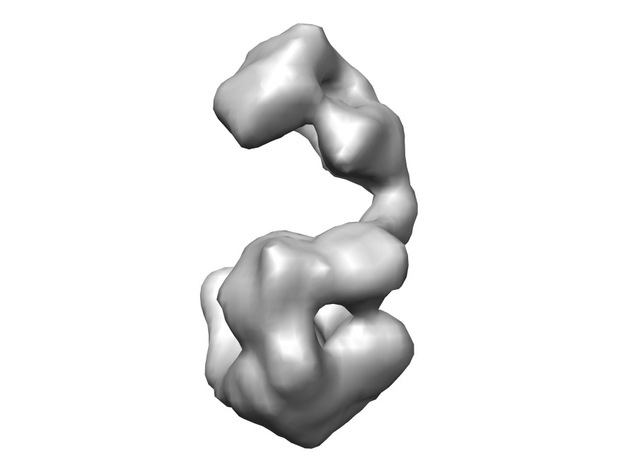
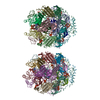
| Title | 3D reconstruction of heterodimeric yeast Pol alpha using electron microscopy | |||||||||
 Map data Map data | 3D reconstruction of heterodimeric yeast Pol alpha using electron microscopy | |||||||||
 Sample Sample |
| |||||||||
 Keywords Keywords | DNA replication / DNA Polymerase alpha / Electron microscopy | |||||||||
| Function / homology |  Function and homology information Function and homology informationprotein binding / RNA-templated DNA biosynthetic process / nucleoside binding / premeiotic DNA replication / alpha DNA polymerase:primase complex / lagging strand elongation / telomere capping / DNA synthesis involved in DNA repair / DNA replication initiation / nuclear envelope ...protein binding / RNA-templated DNA biosynthetic process / nucleoside binding / premeiotic DNA replication / alpha DNA polymerase:primase complex / lagging strand elongation / telomere capping / DNA synthesis involved in DNA repair / DNA replication initiation / nuclear envelope / DNA-directed DNA polymerase activity / nucleotide binding / mitochondrion / DNA binding Similarity search - Function | |||||||||
| Biological species |  | |||||||||
| Method | single particle reconstruction / negative staining / Resolution: 22.9 Å | |||||||||
 Authors Authors | Klinge S / Nunez-Ramirez R / Llorca O / Pellegrini L | |||||||||
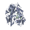
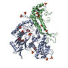
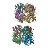
 Citation Citation | Journal: EMBO J / Year: 2009 Title: 3D architecture of DNA Pol alpha reveals the functional core of multi-subunit replicative polymerases. Authors: Sebastian Klinge / Rafael Núñez-Ramírez / Oscar Llorca / Luca Pellegrini /  Abstract: Eukaryotic DNA replication requires the coordinated activity of the multi-subunit DNA polymerases: Pol alpha, Pol delta and Pol epsilon. The conserved catalytic and regulatory B subunits associate in ...Eukaryotic DNA replication requires the coordinated activity of the multi-subunit DNA polymerases: Pol alpha, Pol delta and Pol epsilon. The conserved catalytic and regulatory B subunits associate in a constitutive heterodimer that represents the functional core of all three replicative polymerases. Here, we combine X-ray crystallography and electron microscopy (EM) to describe subunit interaction and 3D architecture of heterodimeric yeast Pol alpha. The crystal structure of the C-terminal domain (CTD) of the catalytic subunit bound to the B subunit illustrates a conserved mechanism of accessory factor recruitment by replicative polymerases. The EM reconstructions of Pol alpha reveal a bilobal shape with separate catalytic and regulatory modules. Docking of the B-CTD complex in the EM reconstruction shows that the B subunit is tethered to the polymerase domain through a structured but flexible linker. Our combined findings provide a structural template for the common functional architecture of the three major replicative DNA polymerases. | |||||||||
| History |
|
- Structure visualization
| Structure viewer | EM map: SurfViewMolmilJmol/JSmol |
|---|---|
| Supplemental images |

- Downloads & links
-EMDB archive
| Map data | emd_1618.map.gz | 68.5 KB | EMDB map data format | |
|---|---|---|---|---|
| Header (meta data) | emd-1618-v30.xmlemd-1618.xml | 13.7 KB 13.7 KB | Display Display | EMDB header |
| Images | EMD-1618.tifemd_1618.tif | 1.6 MB 1.6 MB | ||
| Archive directory |  http://ftp.pdbj.org/pub/emdb/structures/EMD-1618ftp://ftp.pdbj.org/pub/emdb/structures/EMD-1618 http://ftp.pdbj.org/pub/emdb/structures/EMD-1618ftp://ftp.pdbj.org/pub/emdb/structures/EMD-1618 | HTTPS FTP |
-Related structure data


-Links
| EMDB pages | EMDB (EBI/PDBe) / EMDataResource |
|---|
-Map
| File | Download / File: emd_1618.map.gz / Format: CCP4 / Size: 1001 KB / Type: IMAGE STORED AS FLOATING POINT NUMBER (4 BYTES) | ||||||||||||||||||||||||||||||||||||
|---|---|---|---|---|---|---|---|---|---|---|---|---|---|---|---|---|---|---|---|---|---|---|---|---|---|---|---|---|---|---|---|---|---|---|---|---|---|
| Annotation | 3D reconstruction of heterodimeric yeast Pol alpha using electron microscopy | ||||||||||||||||||||||||||||||||||||
| Projections & slices | Image control
Images are generated by Spider. | ||||||||||||||||||||||||||||||||||||
| Voxel size | X=Y=Z: 3.8 Å | ||||||||||||||||||||||||||||||||||||
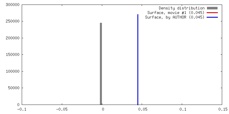
| Density |
| ||||||||||||||||||||||||||||||||||||
| Symmetry | Space group: 1 | ||||||||||||||||||||||||||||||||||||
| Details | EMDB XML:
|
 Z (Sec.)
 Y (Row.)
 X (Col.)


-Supplemental data
- Sample components
-Entire : Yeast DNA polymerase alpha
| Entire | Name: Yeast DNA polymerase alpha |
|---|---|
| Components |
|
-Supramolecule #1000: Yeast DNA polymerase alpha
| Supramolecule | Name: Yeast DNA polymerase alpha / type: sample / ID: 1000 Oligomeric state: One monomer of DNA polymerase alpha binds to a monomer of B-subunit Number unique components: 2 |
|---|---|
| Molecular weight | Theoretical: 185 KDa / Method: Gel Filtration |
-Macromolecule #1: DNA polymerase alpha
| Macromolecule | Name: DNA polymerase alpha / type: protein_or_peptide / ID: 1 / Name.synonym: DNA polymerase alpha / Number of copies: 1 / Oligomeric state: Dimer / Recombinant expression: Yes |
|---|---|
| Source (natural) | Organism: |
| Molecular weight | Experimental: 130 KDa / Theoretical: 130 KDa |
| Sequence | GO: alpha DNA polymerase:primase complex, mitochondrion, DNA binding, DNA-directed DNA polymerase activity, nucleoside binding, nucleotide binding, protein binding, DNA replication initiation, DNA ...GO: alpha DNA polymerase:primase complex, mitochondrion, DNA binding, DNA-directed DNA polymerase activity, nucleoside binding, nucleotide binding, protein binding, DNA replication initiation, DNA synthesis involved in DNA repair, lagging strand elongation, premeiotic DNA replication, RNA-templated DNA biosynthetic process InterPro: DNA-directed DNA polymerase, family B, DNA-directed DNA polymerase, family B, multifunctional domain, INTERPRO: IPR017966, DNA-directed DNA polymerase, family B, conserved site, DNA- ...InterPro: DNA-directed DNA polymerase, family B, DNA-directed DNA polymerase, family B, multifunctional domain, INTERPRO: IPR017966, DNA-directed DNA polymerase, family B, conserved site, DNA-directed DNA polymerase, family B, exonuclease domain, INTERPRO: IPR004578, Zinc finger, DNA-directed DNA polymerase, family B, alpha |
-Macromolecule #2: B subunit
| Macromolecule | Name: B subunit / type: protein_or_peptide / ID: 2 / Name.synonym: B subunit / Number of copies: 1 / Oligomeric state: Dimer / Recombinant expression: Yes |
|---|---|
| Source (natural) | Organism: |
| Molecular weight | Experimental: 55 KDa / Theoretical: 55 KDa |
| Sequence | GO: alpha DNA polymerase:primase complex, nuclear envelope, DNA binding, DNA-directed DNA polymerase activity, protein binding, DNA replication initiation, lagging strand elongation, telomere capping InterPro: DNA polymerase alpha/delta/epsilon, subunit B, DNA polymerase alpha, subunit B, DNA polymerase alpha, subunit B, N-terminal |
-Experimental details
-Structure determination
| Method | negative staining |
|---|---|
 Processing Processing | single particle reconstruction |
| Aggregation state | particle |
-Sample preparation
| Concentration | 0.7 mg/mL |
|---|---|
| Buffer | pH: 8 / Details: 100mM TrisHCl pH 8.0, 500 mM NaCl |
| Staining | Type: NEGATIVE Details: A diluted solution of Pol alpha complex was adsorbed onto glow-discharged carbon coated grids, stained with 2% uranyl formate |
| Grid | Details: 400 mesh copper grid |
| Vitrification | Cryogen name: NONE / Instrument: OTHER |
- Electron microscopy
| Microscope | JEOL 1230 |
|---|---|
| Temperature | Min: 293 K / Max: 293 K / Average: 293 K |
| Alignment procedure | Legacy - Astigmatism: Objective lens astigmatism was corrected at 120,000 times magnification |
| Details | Microscope used JEOL-1230 |
| Date | Aug 20, 2009 |
| Image recording | Category: FILM / Film or detector model: KODAK SO-163 FILM / Digitization - Scanner: OTHER / Digitization - Sampling interval: 10 µm / Number real images: 170 / Average electron dose: 15 e/Å2 / Bits/pixel: 8 |
| Electron beam | Acceleration voltage: 100 kV / Electron source: TUNGSTEN HAIRPIN |
| Electron optics | Illumination mode: FLOOD BEAM / Imaging mode: BRIGHT FIELD / Cs: 2.9 mm / Nominal defocus max: 1.2 µm / Nominal defocus min: 0.75 µm / Nominal magnification: 50000 |
| Sample stage | Specimen holder: Eucentric / Specimen holder model: JEOL |
-Image processing
| CTF correction | Details: reverse phases |
|---|---|
| Final reconstruction | Applied symmetry - Point group: C1 (asymmetric) / Algorithm: OTHER / Resolution.type: BY AUTHOR / Resolution: 22.9 Å / Resolution method: FSC 0.5 CUT-OFF / Software - Name: EMAN, Xmipp / Number images used: 12913 |
| Final two d classification | Number classes: 190 |
-Atomic model buiding 1
| Initial model | PDB ID: |
|---|---|
| Software | Name: adpem |
| Details | Protocol: Rigid Body |
| Refinement | Space: REAL / Protocol: RIGID BODY FIT / Target criteria: cross-correlation |
-Atomic model buiding 2
| Initial model | PDB ID: |
|---|---|
| Software | Name: adpem |
| Details | Protocol: Rigid Body |
| Refinement | Space: REAL / Protocol: RIGID BODY FIT / Target criteria: cross-correlation |