 Movie
Movie Controller

[English] 日本語
 Yorodumi
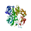
- PDB-2vwj: Uracil Recognition in Archaeal DNA Polymerases Captured by X-ray ... -
+ Open data

- Basic information
| Entry | Database: PDB / ID: 2vwj | ||||||
|---|---|---|---|---|---|---|---|
| Title | Uracil Recognition in Archaeal DNA Polymerases Captured by X-ray Crystallography. | ||||||
 Components Components |
| ||||||
 Keywords Keywords | DNA REPLICATION / MULTIFUNCTIONAL ENZYME / NUCLEOTIDYLTRANSFERASE / DNA-DIRECTED DNA POLYMERASE / TRANSFERASE / EXONUCLEASE / DNA-BINDING / DNA POLYMERASE / URACIL / ARCHAEA / NUCLEASE / HYDROLASE / DNA REPAIR | ||||||
| Function / homology |  Function and homology information Function and homology informationexonuclease activity / DNA-templated DNA replication / DNA-directed DNA polymerase / DNA-directed DNA polymerase activity / nucleotide binding / DNA binding Similarity search - Function | ||||||
| Biological species |   THERMOCOCCUS GORGONARIUS (archaea) THERMOCOCCUS GORGONARIUS (archaea) | ||||||
| Method |  X-RAY DIFFRACTION / SYNCHROTRON / MOLECULAR REPLACEMENT / Resolution: 2.78 Å X-RAY DIFFRACTION / SYNCHROTRON / MOLECULAR REPLACEMENT / Resolution: 2.78 Å | ||||||
 Authors Authors | Firbank, S.J. / Wardle, J. / Heslop, P. / Lewis, R.J. / Connolly, B.A. | ||||||
 Citation Citation | Journal: J.Mol.Biol. / Year: 2008 Title: Uracil Recognition in Archaeal DNA Polymerases Captured by X-Ray Crystallography. Authors: Firbank, S.J. / Wardle, J. / Heslop, P. / Lewis, R.J. / Connolly, B.A. | ||||||
| History |
|
- Structure visualization
| Structure viewer | Molecule: MolmilJmol/JSmol |
|---|
- Downloads & links
-Download
| PDBx/mmCIF format | 2vwj.cif.gz | 179.1 KB | Display | PDBx/mmCIF format |
|---|---|---|---|---|
| PDB format | pdb2vwj.ent.gz | 136.3 KB | Display | PDB format |
| PDBx/mmJSON format | 2vwj.json.gz | Tree view | PDBx/mmJSON format | |
| Others |  Other downloads Other downloads |
-Validation report
| Arichive directory | https://data.pdbj.org/pub/pdb/validation_reports/vw/2vwjftp://data.pdbj.org/pub/pdb/validation_reports/vw/2vwj | HTTPS FTP |
|---|
-Related structure data
| Related structure data | 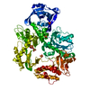 2vwkC  1tgoS S: Starting model for refinement C: citing same article ( |
|---|---|
| Similar structure data |


-Links
 PDBj
PDBj


- Assembly
| Deposited unit | 
| ||||||||
|---|---|---|---|---|---|---|---|---|---|
| 1 |
| ||||||||
| Unit cell |
|
-Components
| #1: Protein | Mass: 89936.305 Da / Num. of mol.: 1 / Mutation: YES Source method: isolated from a genetically manipulated source Source: (gene. exp.) THERMOCOCCUS GORGONARIUS (archaea) / Plasmid: PET / Production host:  | ||||||
|---|---|---|---|---|---|---|---|
| #2: DNA chain | Mass: 8005.138 Da / Num. of mol.: 1 / Source method: obtained synthetically | ||||||
| #3: Chemical |   Mass: 39.098 Da / Num. of mol.: 3 / Source method: obtained synthetically / Formula: K Mass: 39.098 Da / Num. of mol.: 3 / Source method: obtained synthetically / Formula: K#4: Water | ChemComp-HOH / |  Mass: 18.015 Da / Num. of mol.: 1 / Source method: isolated from a natural source / Formula: H2O Mass: 18.015 Da / Num. of mol.: 1 / Source method: isolated from a natural source / Formula: H2OCompound details | ENGINEERED | Sequence details | MUTATION OF D215 TO ALANINE TO REMOVE EXONUCLEAS | |
-Experimental details
-Experiment
| Experiment | Method: X-RAY DIFFRACTION / Number of used crystals: 1 |
|---|
- Sample preparation
| Crystal | Density Matthews: 4.5 Å3/Da / Density % sol: 76 % / Description: NONE |
|---|---|
| Crystal grow | pH: 8.3 / Details: 0.2M TRIPOTASSIUM CITRATE PH8.3, 20% PEG 3350 |
-Data collection
| Diffraction | Mean temperature: 100 K |
|---|---|
| Diffraction source | Source: SYNCHROTRON / Site: Diamond  / Beamline: I04 / Wavelength: 0.9699 / Beamline: I04 / Wavelength: 0.9699 |
| Detector | Type: ADSC QUANTUM 315 / Detector: CCD Details: SI (111) DOUBLE CRYSTAL MONOCHROMATOR. KIRKPATRICK BAEZ BIMORPH MIRROR PAIR FOR HORIZONTAL AND VERTICAL FOCUSSING |
| Radiation | Monochromator: DOUBLE CRYSTAL / Protocol: SINGLE WAVELENGTH / Monochromatic (M) / Laue (L): M / Scattering type: x-ray |
| Radiation wavelength | Wavelength: 0.9699 Å / Relative weight: 1 |
| Reflection | Resolution: 2.78→30 Å / Num. obs: 45193 / % possible obs: 98.7 % / Observed criterion σ(I): 0 / Redundancy: 3.5 % / Rmerge(I) obs: 0.1 / Net I/σ(I): 11.1 |
| Reflection shell | Resolution: 2.78→2.85 Å / Redundancy: 3.6 % / Rmerge(I) obs: 0.44 / Mean I/σ(I) obs: 2.2 / % possible all: 98.9 |
- Processing
| Software |
| ||||||||||||||||||||||||||||||||||||||||||||||||||||||||||||||||||||||||||||||||||||||||||||||||||||||||||||||||||||||||||||||||||||||||||||||||||||||||||||||||||||||||||||||||||||||
|---|---|---|---|---|---|---|---|---|---|---|---|---|---|---|---|---|---|---|---|---|---|---|---|---|---|---|---|---|---|---|---|---|---|---|---|---|---|---|---|---|---|---|---|---|---|---|---|---|---|---|---|---|---|---|---|---|---|---|---|---|---|---|---|---|---|---|---|---|---|---|---|---|---|---|---|---|---|---|---|---|---|---|---|---|---|---|---|---|---|---|---|---|---|---|---|---|---|---|---|---|---|---|---|---|---|---|---|---|---|---|---|---|---|---|---|---|---|---|---|---|---|---|---|---|---|---|---|---|---|---|---|---|---|---|---|---|---|---|---|---|---|---|---|---|---|---|---|---|---|---|---|---|---|---|---|---|---|---|---|---|---|---|---|---|---|---|---|---|---|---|---|---|---|---|---|---|---|---|---|---|---|---|---|
| Refinement | Method to determine structure: MOLECULAR REPLACEMENT Starting model: PDB ENTRY 1TGO Resolution: 2.78→67.73 Å / Cor.coef. Fo:Fc: 0.914 / Cor.coef. Fo:Fc free: 0.889 / SU B: 11.645 / SU ML: 0.228 / Cross valid method: THROUGHOUT / ESU R: 0.405 / ESU R Free: 0.294 / Stereochemistry target values: MAXIMUM LIKELIHOOD / Details: HYDROGENS HAVE BEEN ADDED IN THE RIDING POSITIONS.
| ||||||||||||||||||||||||||||||||||||||||||||||||||||||||||||||||||||||||||||||||||||||||||||||||||||||||||||||||||||||||||||||||||||||||||||||||||||||||||||||||||||||||||||||||||||||
| Solvent computation | Ion probe radii: 0.8 Å / Shrinkage radii: 0.8 Å / VDW probe radii: 1.2 Å / Solvent model: MASK | ||||||||||||||||||||||||||||||||||||||||||||||||||||||||||||||||||||||||||||||||||||||||||||||||||||||||||||||||||||||||||||||||||||||||||||||||||||||||||||||||||||||||||||||||||||||
| Displacement parameters | Biso mean: 52.83 Å2
| ||||||||||||||||||||||||||||||||||||||||||||||||||||||||||||||||||||||||||||||||||||||||||||||||||||||||||||||||||||||||||||||||||||||||||||||||||||||||||||||||||||||||||||||||||||||
| Refinement step | Cycle: LAST / Resolution: 2.78→67.73 Å
| ||||||||||||||||||||||||||||||||||||||||||||||||||||||||||||||||||||||||||||||||||||||||||||||||||||||||||||||||||||||||||||||||||||||||||||||||||||||||||||||||||||||||||||||||||||||
| Refine LS restraints |
|