 Movie
Movie Controller

[English] 日本語
 Yorodumi
- EMDB-15442: In situ cryo-electron tomogram of an intact pluripotent P19 cellu... -
+ Open data

- Basic information
| Entry |  | |||||||||
|---|---|---|---|---|---|---|---|---|---|---|
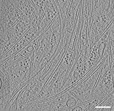
| Title | In situ cryo-electron tomogram of an intact pluripotent P19 cellular process after nocodazole treatment and washout. | |||||||||
 Map data Map data | In situ cryo-electron tomogram of an intact pluripotent P19 cellular process after nocodazole treatment and washout. | |||||||||
 Sample Sample |
| |||||||||
 Keywords Keywords | Microtubules / Scaffold / protein transport | |||||||||
| Biological species |  | |||||||||
| Method | electron tomography / cryo EM | |||||||||
 Authors Authors | Chakraborty S / Martinez-Sanchez A / Baumeister W / Mahamid J | |||||||||
| Funding support |  Germany, 1 items Germany, 1 items
| |||||||||
 Citation Citation | Journal: Biorxiv / Year: 2024 Title: Cryo-electron tomography suggests tubulin chaperones form a subset of microtubule lumenal particles with a role in maintaining neuronal microtubules Authors: Chakraborty S / Martinez-Sanchez A / Beck F / Toro-Nahuelpan M / Hwang IY / Noh KM / Baumeister W / Mahamid J | |||||||||
| History |
|
- Structure visualization
| Supplemental images |
|---|
- Downloads & links
-EMDB archive
| Map data | emd_15442.map.gz | 190.3 MB |  EMDB map data format EMDB map data format | |
|---|---|---|---|---|
| Header (meta data) | emd-15442-v30.xmlemd-15442.xml | 9.4 KB 9.4 KB | Display Display | EMDB header |
| Images |  emd_15442.png emd_15442.png | 251.7 KB | ||
| Filedesc metadata | emd-15442.cif.gz | 3.9 KB | ||
| Archive directory |  http://ftp.pdbj.org/pub/emdb/structures/EMD-15442ftp://ftp.pdbj.org/pub/emdb/structures/EMD-15442 http://ftp.pdbj.org/pub/emdb/structures/EMD-15442ftp://ftp.pdbj.org/pub/emdb/structures/EMD-15442 | HTTPS FTP |
-Validation report
| Summary document | emd_15442_validation.pdf.gz | 649.7 KB | Display | EMDB validaton report |
|---|---|---|---|---|
| Full document | emd_15442_full_validation.pdf.gz | 649.2 KB | Display | |
| Data in XML | emd_15442_validation.xml.gz | 4.8 KB | Display | |
| Data in CIF | emd_15442_validation.cif.gz | 5.3 KB | Display | |
| Arichive directory | https://ftp.pdbj.org/pub/emdb/validation_reports/EMD-15442ftp://ftp.pdbj.org/pub/emdb/validation_reports/EMD-15442 | HTTPS FTP |
-Related structure data
| Related structure data | C: citing same article ( |
|---|


-Links
| EMDB pages | EMDB (EBI/PDBe) / EMDataResource |
|---|
-Map
| File | Download / File: emd_15442.map.gz / Format: CCP4 / Size: 205.3 MB / Type: IMAGE STORED AS FLOATING POINT NUMBER (4 BYTES) | ||||||||||||||||||||||||||||||||
|---|---|---|---|---|---|---|---|---|---|---|---|---|---|---|---|---|---|---|---|---|---|---|---|---|---|---|---|---|---|---|---|---|---|
| Annotation | In situ cryo-electron tomogram of an intact pluripotent P19 cellular process after nocodazole treatment and washout. | ||||||||||||||||||||||||||||||||
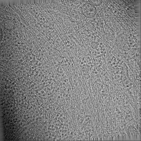
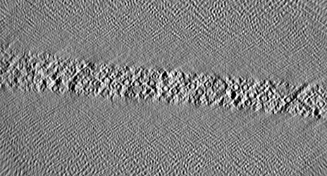
| Projections & slices | Image control
Images are generated by Spider. generated in cubic-lattice coordinate | ||||||||||||||||||||||||||||||||
| Voxel size | X=Y=Z: 27.36 Å | ||||||||||||||||||||||||||||||||
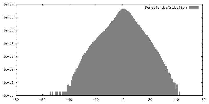
| Density |
| ||||||||||||||||||||||||||||||||
| Symmetry | Space group: 1 | ||||||||||||||||||||||||||||||||
| Details | EMDB XML:
|
 Z (Sec.)
 Y (Row.)
 X (Col.)


-Supplemental data
- Sample components
-Entire : In situ cryo-electron tomogram of an intact pluripotent P19 cellu...
| Entire | Name: In situ cryo-electron tomogram of an intact pluripotent P19 cellular process after nocodazole treatment and washout. |
|---|---|
| Components |
|
-Supramolecule #1: In situ cryo-electron tomogram of an intact pluripotent P19 cellu...
| Supramolecule | Name: In situ cryo-electron tomogram of an intact pluripotent P19 cellular process after nocodazole treatment and washout. type: cell / ID: 1 / Parent: 0 Details: The cells were treated with nocodazole for 30 minutes followed by washing out the drug. |
|---|---|
| Source (natural) | Organism: |
-Experimental details
-Structure determination
| Method | cryo EM |
|---|---|
 Processing Processing | electron tomography |
| Aggregation state | cell |
-Sample preparation
| Buffer | pH: 7.2 |
|---|---|
| Grid | Model: Quantifoil R2/1 / Material: GOLD / Mesh: 200 / Pretreatment - Type: GLOW DISCHARGE / Pretreatment - Time: 45 sec. |
| Vitrification | Cryogen name: ETHANE-PROPANE / Chamber humidity: 95 % / Chamber temperature: 310 K / Instrument: FEI VITROBOT MARK IV Details: Blotted from backside for 10 seconds with blot force 10. |
| Sectioning | Other: NO SECTIONING |
- Electron microscopy
| Microscope | TFS KRIOS |
|---|---|
| Temperature | Min: 77.0 K |
| Specialist optics | Phase plate: VOLTA PHASE PLATE / Energy filter - Name: GIF Quantum LS / Energy filter - Slit width: 20 eV |
| Image recording | Film or detector model: GATAN K2 SUMMIT (4k x 4k) / Detector mode: COUNTING / Average electron dose: 1.4 e/Å2 |
| Electron beam | Acceleration voltage: 300 kV / Electron source:  FIELD EMISSION GUN FIELD EMISSION GUN |
| Electron optics | C2 aperture diameter: 70.0 µm / Illumination mode: SPOT SCAN / Imaging mode: BRIGHT FIELD / Cs: 2.7 mm / Nominal defocus max: 0.5 µm / Nominal defocus min: 0.2 µm / Nominal magnification: 42000 |
| Sample stage | Specimen holder model: FEI TITAN KRIOS AUTOGRID HOLDER / Cooling holder cryogen: NITROGEN |
| Experimental equipment |  Model: Titan Krios / Image courtesy: FEI Company |
-Image processing
| Final reconstruction | Algorithm: BACK PROJECTION / Software - Name: IMOD (ver. 4.9.0) / Number images used: 61 |
|---|
