 Movie
Movie Controller

[English] 日本語
 Yorodumi
- EMDB-15468: In situ subtomogram average of floating microtubule inner protein... -
+ Open data

- Basic information
| Entry |  | |||||||||
|---|---|---|---|---|---|---|---|---|---|---|
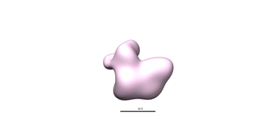
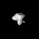
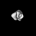
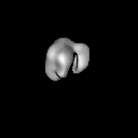
| Title | In situ subtomogram average of floating microtubule inner protein 16 (fMIP16) | |||||||||
 Map data Map data | In situ subtomogram average of floating microtubule inner protein 16 | |||||||||
 Sample Sample |
| |||||||||
 Keywords Keywords | microtubule associated protein / STRUCTURAL PROTEIN | |||||||||
| Biological species |  Homo sapiens (human) Homo sapiens (human) | |||||||||
| Method | subtomogram averaging / cryo EM / Resolution: 27.0 Å | |||||||||
 Authors Authors | Chakraborty S / Mahamid J / Martinez-Sanchez A / Baumeister W | |||||||||
| Funding support | 1 items
| |||||||||
 Citation Citation | Journal: Proc Natl Acad Sci U S A / Year: 2025 Title: Cryo-ET suggests tubulin chaperones form a subset of microtubule lumenal particles with a role in maintaining neuronal microtubules. Authors: Saikat Chakraborty / Antonio Martinez-Sanchez / Florian Beck / Mauricio Toro-Nahuelpan / In-Young Hwang / Kyung-Min Noh / Wolfgang Baumeister / Julia Mahamid /  Abstract: The functional architecture of the long-lived neuronal microtubule (MT) cytoskeleton is maintained by various MT-associated proteins (MAPs), most of which are known to bind to the MT outer surface. ...The functional architecture of the long-lived neuronal microtubule (MT) cytoskeleton is maintained by various MT-associated proteins (MAPs), most of which are known to bind to the MT outer surface. However, electron microscopy (EM) has long ago revealed the presence of particles inside the lumens of neuronal MTs, of yet unknown identity and function. Here, we use cryogenic electron tomography (cryo-ET) to analyze the three-dimensional (3D) organization and structures of MT lumenal particles in primary hippocampal neurons, human induced pluripotent stem cell-derived neurons, and pluripotent and differentiated P19 cells. We obtain in situ density maps of several lumenal particles from the respective cells and detect common structural features underscoring their potential overarching functions. Mass spectrometry-based proteomics combined with structural modeling suggest that a subset of lumenal particles could be tubulin-binding cofactors (TBCs) bound to tubulin monomers. A different subset of smaller particles, which remains unidentified, exhibits densities that bridge across the MT protofilaments. We show that increased lumenal particle concentration within MTs is concomitant with neuronal differentiation and correlates with higher MT curvatures. Enrichment of lumenal particles around MT lattice defects and at freshly polymerized MT open-ends suggests a MT protective role. Together with the identified structural resemblance of a subset of particles to TBCs, these results hint at a role in local tubulin proteostasis for the maintenance of long-lived neuronal MTs. #1: Journal: Biorxiv / Year: 2024Title: Cryo-electron tomography suggests tubulin chaperones form a subset of microtubule lumenal particles with a role in maintaining neuronal microtubules Authors: Chakraborty S / Martinez-Sanchez A / Beck F / Toro-Nahuelpan M / Hwang IY / Noh KM / Baumeister W / Mahamid J | |||||||||
| History |
|
- Structure visualization
| Supplemental images |
|---|
- Downloads & links
-EMDB archive
| Map data | emd_15468.map.gz | 2.1 MB |  EMDB map data format EMDB map data format | |
|---|---|---|---|---|
| Header (meta data) | emd-15468-v30.xmlemd-15468.xml | 16.6 KB 16.6 KB | Display Display | EMDB header |
| FSC (resolution estimation) | emd_15468_fsc.xml | 4.7 KB | Display | FSC data file |
| Images |  emd_15468.png emd_15468.png | 9.3 KB | ||
| Filedesc metadata | emd-15468.cif.gz | 4.3 KB | ||
| Others | emd_15468_half_map_1.map.gzemd_15468_half_map_2.map.gz | 6 MB 6 MB | ||
| Archive directory |  http://ftp.pdbj.org/pub/emdb/structures/EMD-15468ftp://ftp.pdbj.org/pub/emdb/structures/EMD-15468 http://ftp.pdbj.org/pub/emdb/structures/EMD-15468ftp://ftp.pdbj.org/pub/emdb/structures/EMD-15468 | HTTPS FTP |
-Related structure data
| Related structure data | C: citing same article ( |
|---|


-Links
| EMDB pages | EMDB (EBI/PDBe) / EMDataResource |
|---|
-Map
| File | Download / File: emd_15468.map.gz / Format: CCP4 / Size: 8 MB / Type: IMAGE STORED AS FLOATING POINT NUMBER (4 BYTES) | ||||||||||||||||||||||||||||||||||||
|---|---|---|---|---|---|---|---|---|---|---|---|---|---|---|---|---|---|---|---|---|---|---|---|---|---|---|---|---|---|---|---|---|---|---|---|---|---|
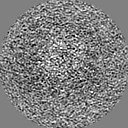
| Annotation | In situ subtomogram average of floating microtubule inner protein 16 | ||||||||||||||||||||||||||||||||||||
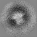
| Projections & slices | Image control
Images are generated by Spider. | ||||||||||||||||||||||||||||||||||||
| Voxel size | X=Y=Z: 2.129 Å | ||||||||||||||||||||||||||||||||||||
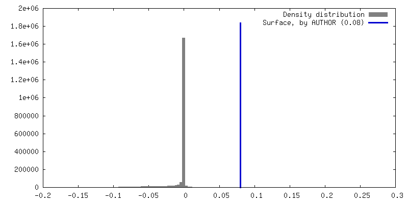
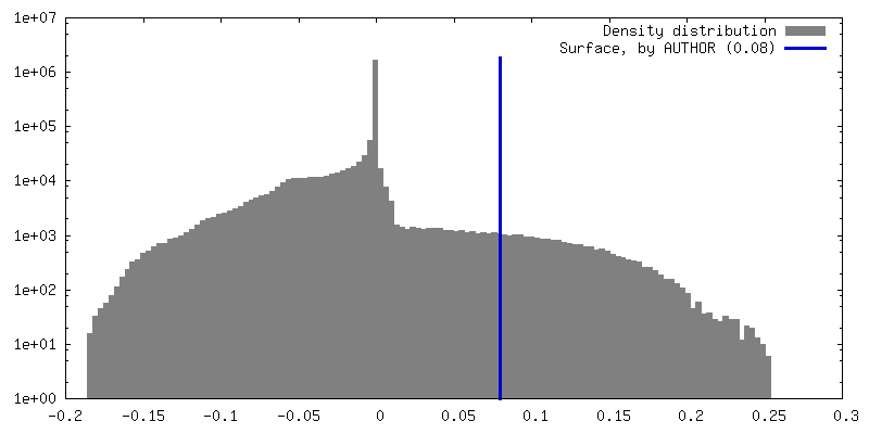
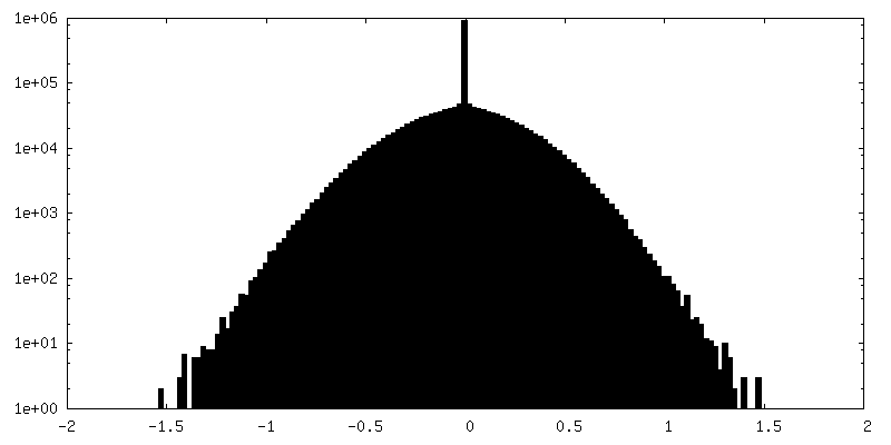
| Density |
| ||||||||||||||||||||||||||||||||||||
| Symmetry | Space group: 1 | ||||||||||||||||||||||||||||||||||||
| Details | EMDB XML:
|
 Z (Sec.)
 Y (Row.)
 X (Col.)


-Supplemental data
-Half map: In situ subtomogram average of floating microtubule inner protein 16
| File | emd_15468_half_map_1.map | ||||||||||||
|---|---|---|---|---|---|---|---|---|---|---|---|---|---|
| Annotation | In situ subtomogram average of floating microtubule inner protein 16 | ||||||||||||
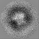
| Projections & Slices |
| ||||||||||||
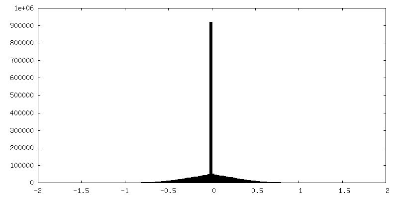
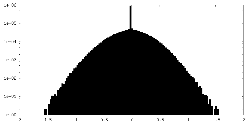
| Density Histograms |
-Half map: In situ subtomogram average of floating microtubule inner protein 16
| File | emd_15468_half_map_2.map | ||||||||||||
|---|---|---|---|---|---|---|---|---|---|---|---|---|---|
| Annotation | In situ subtomogram average of floating microtubule inner protein 16 | ||||||||||||
| Projections & Slices |
| ||||||||||||
| Density Histograms |

- Sample components
-Entire : fMIP16 from human induced pluripotent stem cells derived neuronal...
| Entire | Name: fMIP16 from human induced pluripotent stem cells derived neuronal microtubules |
|---|---|
| Components |
|
-Supramolecule #1: fMIP16 from human induced pluripotent stem cells derived neuronal...
| Supramolecule | Name: fMIP16 from human induced pluripotent stem cells derived neuronal microtubules type: cell / ID: 1 / Parent: 0 |
|---|---|
| Source (natural) | Organism: Homo sapiens (human) |
-Experimental details
-Structure determination
| Method | cryo EM |
|---|---|
 Processing Processing | subtomogram averaging |
| Aggregation state | cell |
-Sample preparation
| Buffer | pH: 7.2 |
|---|---|
| Grid | Model: Quantifoil / Material: GOLD / Mesh: 200 / Pretreatment - Type: GLOW DISCHARGE / Pretreatment - Time: 30 sec. |
| Vitrification | Cryogen name: ETHANE-PROPANE / Chamber humidity: 70 % / Chamber temperature: 310 K / Instrument: LEICA EM GP |
| Details | Frozen hydrated human induced pluripotent stem cells derived neurons |
- Electron microscopy
| Microscope | FEI TITAN KRIOS |
|---|---|
| Temperature | Min: 77.0 K |
| Specialist optics | Phase plate: VOLTA PHASE PLATE / Energy filter - Name: GIF Quantum LS / Energy filter - Slit width: 20 eV |
| Image recording | Film or detector model: GATAN K2 SUMMIT (4k x 4k) / Detector mode: COUNTING / Average electron dose: 2.0 e/Å2 |
| Electron beam | Acceleration voltage: 300 kV / Electron source:  FIELD EMISSION GUN FIELD EMISSION GUN |
| Electron optics | C2 aperture diameter: 70.0 µm / Illumination mode: FLOOD BEAM / Imaging mode: BRIGHT FIELD / Cs: 2.7 mm / Nominal defocus max: 4.0 µm / Nominal defocus min: 2.5 µm / Nominal magnification: 64000 |
| Sample stage | Specimen holder model: FEI TITAN KRIOS AUTOGRID HOLDER / Cooling holder cryogen: NITROGEN |
| Experimental equipment |  Model: Titan Krios / Image courtesy: FEI Company |

