 Movie
Movie Controller

[English] 日本語
 Yorodumi
- EMDB-14491: Assymetric reconstruction of bacteriophage SU10 genome release in... -
+ Open data

- Basic information
| Entry |  | |||||||||
|---|---|---|---|---|---|---|---|---|---|---|
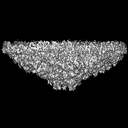
| Title | Assymetric reconstruction of bacteriophage SU10 genome release intermediate capsid cap | |||||||||
 Map data Map data | ||||||||||
 Sample Sample |
| |||||||||
 Keywords Keywords | bacteriophage / capsid / assymetric / VIRUS | |||||||||
| Biological species |  Escherichia phage vB_EcoP_SU10 (virus) Escherichia phage vB_EcoP_SU10 (virus) | |||||||||
| Method | single particle reconstruction / cryo EM / Resolution: 7.3 Å | |||||||||
 Authors Authors | Siborova M / Fuzik T / Prochazkova M / Novacek J / Plevka P | |||||||||
| Funding support |  Czech Republic, 1 items Czech Republic, 1 items
| |||||||||
 Citation Citation | Journal: Nat Commun / Year: 2022 Title: Tail proteins of phage SU10 reorganize into the nozzle for genome delivery. Authors: Marta Šiborová / Tibor Füzik / Michaela Procházková / Jiří Nováček / Martin Benešík / Anders S Nilsson / Pavel Plevka /  Abstract: Escherichia coli phage SU10 belongs to the genus Kuravirus from the class Caudoviricetes of phages with short non-contractile tails. In contrast to other short-tailed phages, the tails of Kuraviruses ...Escherichia coli phage SU10 belongs to the genus Kuravirus from the class Caudoviricetes of phages with short non-contractile tails. In contrast to other short-tailed phages, the tails of Kuraviruses elongate upon cell attachment. Here we show that the virion of SU10 has a prolate head, containing genome and ejection proteins, and a tail, which is formed of portal, adaptor, nozzle, and tail needle proteins and decorated with long and short fibers. The binding of the long tail fibers to the receptors in the outer bacterial membrane induces the straightening of nozzle proteins and rotation of short tail fibers. After the re-arrangement, the nozzle proteins and short tail fibers alternate to form a nozzle that extends the tail by 28 nm. Subsequently, the tail needle detaches from the nozzle proteins and five types of ejection proteins are released from the SU10 head. The nozzle with the putative extension formed by the ejection proteins enables the delivery of the SU10 genome into the bacterial cytoplasm. It is likely that this mechanism of genome delivery, involving the formation of the tail nozzle, is employed by all Kuraviruses. | |||||||||
| History |
|
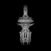
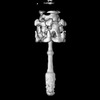
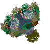
- Structure visualization
| Supplemental images |
|---|

- Downloads & links
-EMDB archive
| Map data | emd_14491.map.gz | 13.1 MB |  EMDB map data format EMDB map data format | |
|---|---|---|---|---|
| Header (meta data) | emd-14491-v30.xmlemd-14491.xml | 20.3 KB 20.3 KB | Display Display | EMDB header |
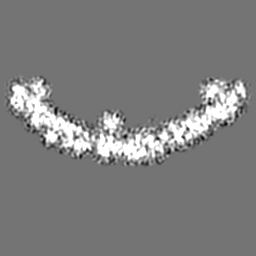
| Images |  emd_14491.png emd_14491.png | 100.1 KB | ||
| Filedesc metadata | emd-14491.cif.gz | 5.5 KB | ||
| Others | emd_14491_half_map_1.map.gzemd_14491_half_map_2.map.gz | 49.6 MB 49.6 MB | ||
| Archive directory |  http://ftp.pdbj.org/pub/emdb/structures/EMD-14491ftp://ftp.pdbj.org/pub/emdb/structures/EMD-14491 http://ftp.pdbj.org/pub/emdb/structures/EMD-14491ftp://ftp.pdbj.org/pub/emdb/structures/EMD-14491 | HTTPS FTP |
-Related structure data


-Links
| EMDB pages | EMDB (EBI/PDBe) / EMDataResource |
|---|
-Map
| File | Download / File: emd_14491.map.gz / Format: CCP4 / Size: 64 MB / Type: IMAGE STORED AS FLOATING POINT NUMBER (4 BYTES) | ||||||||||||||||||||||||||||||||||||
|---|---|---|---|---|---|---|---|---|---|---|---|---|---|---|---|---|---|---|---|---|---|---|---|---|---|---|---|---|---|---|---|---|---|---|---|---|---|
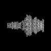
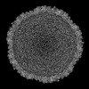
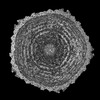
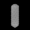
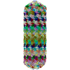
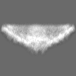
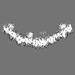
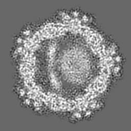
| Projections & slices | Image control
Images are generated by Spider. | ||||||||||||||||||||||||||||||||||||
| Voxel size | X=Y=Z: 1.34 Å | ||||||||||||||||||||||||||||||||||||
| Density |
| ||||||||||||||||||||||||||||||||||||
| Symmetry | Space group: 1 | ||||||||||||||||||||||||||||||||||||
| Details | EMDB XML:
|
 Z (Sec.)
 Y (Row.)
 X (Col.)


-Supplemental data
-Half map: #2
| File | emd_14491_half_map_1.map | ||||||||||||
|---|---|---|---|---|---|---|---|---|---|---|---|---|---|
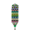
| Projections & Slices |
| ||||||||||||
| Density Histograms |


-Half map: #1
| File | emd_14491_half_map_2.map | ||||||||||||
|---|---|---|---|---|---|---|---|---|---|---|---|---|---|
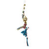
| Projections & Slices |
| ||||||||||||
| Density Histograms |


- Sample components
-Entire : Escherichia phage vB_EcoP_SU10
| Entire | Name: Escherichia phage vB_EcoP_SU10 (virus) |
|---|---|
| Components |
|
-Supramolecule #1: Escherichia phage vB_EcoP_SU10
| Supramolecule | Name: Escherichia phage vB_EcoP_SU10 / type: virus / ID: 1 / Parent: 0 / Macromolecule list: all / NCBI-ID: 1519788 / Sci species name: Escherichia phage vB_EcoP_SU10 / Virus type: VIRION / Virus isolate: STRAIN / Virus enveloped: No / Virus empty: Yes |
|---|---|
| Host (natural) | Organism:  |
| Molecular weight | Theoretical: 3 MDa |
| Virus shell | Shell ID: 1 / Name: Capsid cap assymetric |
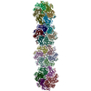
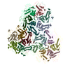
-Macromolecule #1: major capsid protein
| Macromolecule | Name: major capsid protein / type: protein_or_peptide / ID: 1 / Enantiomer: LEVO |
|---|---|
| Source (natural) | Organism: Escherichia phage vB_EcoP_SU10 (virus) |
| Sequence | String: MANPTLFVSY DQNGKKLSFA NWISVLSPQD TPFVSMTGKE SINQ TIFSW QTDALASVDG NNAHVEGSRA EDGEMKPTVI KSNVTQILRK VVRVSDTANT TAN YGRGRE LMYQLEKKGK EIKRDLEKIL LSGQARTDVL ADQYLTNSAA DPAVAGLNDT HA ARKTGAF ...String: MANPTLFVSY DQNGKKLSFA NWISVLSPQD TPFVSMTGKE SINQ TIFSW QTDALASVDG NNAHVEGSRA EDGEMKPTVI KSNVTQILRK VVRVSDTANT TAN YGRGRE LMYQLEKKGK EIKRDLEKIL LSGQARTDVL ADQYLTNSAA DPAVAGLNDT HA ARKTGAF QFLCAHGGLA GGVVDKTKNG PADPDTGAVT VKVAQNASNP TTNIGFDEAD I FDMTLQLY TAGSEADIIM INPAHAKIFA GLQENTQGSR KRIFENTKQF IYEVNSITDP LGQSYKIIV NRWMPTDAVY FFRSADWTQM VLRAPKRTEL AKDGSYEKWM IEMEVGLRH RNPYASGVLF TAAGKAAA GENBANK: GENBANK: NC_027395.1 |
-Experimental details
-Structure determination
| Method | cryo EM |
|---|---|
 Processing Processing | single particle reconstruction |
| Aggregation state | particle |
-Sample preparation
| Buffer | pH: 8 Component:
| ||||||||||||
|---|---|---|---|---|---|---|---|---|---|---|---|---|---|
| Grid | Model: Quantifoil R2/1 / Material: COPPER / Mesh: 200 / Support film - Material: CARBON / Support film - topology: HOLEY / Pretreatment - Type: GLOW DISCHARGE / Pretreatment - Time: 30 sec. / Pretreatment - Atmosphere: OTHER | ||||||||||||
| Vitrification | Cryogen name: ETHANE / Instrument: FEI VITROBOT MARK IV | ||||||||||||
| Details | PFU 10^11 |
- Electron microscopy
| Microscope | FEI TITAN KRIOS |
|---|---|
| Image recording | Film or detector model: GATAN K2 SUMMIT (4k x 4k) / Detector mode: SUPER-RESOLUTION / Number grids imaged: 1 / Average exposure time: 8.0 sec. / Average electron dose: 52.0 e/Å2 |
| Electron beam | Acceleration voltage: 300 kV / Electron source:  FIELD EMISSION GUN FIELD EMISSION GUN |
| Electron optics | C2 aperture diameter: 70.0 µm / Illumination mode: FLOOD BEAM / Imaging mode: BRIGHT FIELD / Cs: 2.7 mm / Nominal defocus max: 2.7 µm / Nominal defocus min: 1.2 µm / Nominal magnification: 10500 |
| Sample stage | Specimen holder model: FEI TITAN KRIOS AUTOGRID HOLDER / Cooling holder cryogen: NITROGEN |
| Experimental equipment |  Model: Titan Krios / Image courtesy: FEI Company |
+Image processing
-Atomic model buiding 1
| Refinement | Space: REAL / Protocol: AB INITIO MODEL |
|---|
