 Movie
Movie Controller

[English] 日本語
 Yorodumi
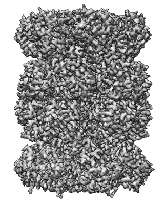
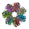
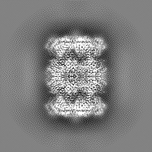
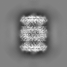
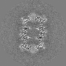
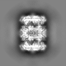
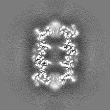
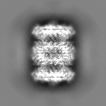
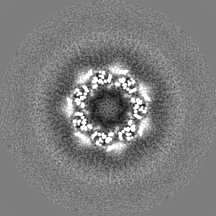
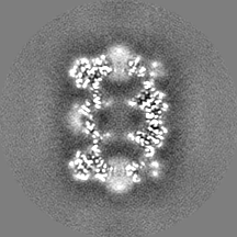
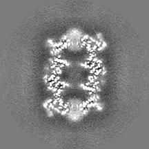
- EMDB-13902: The cryoEM density map of T20s proteasome with various ice thickn... -
+ Open data

- Basic information
| Entry | Database: EMDB / ID: EMD-13902 | |||||||||||||||
|---|---|---|---|---|---|---|---|---|---|---|---|---|---|---|---|---|
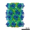
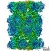
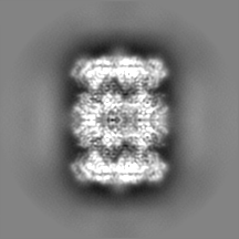
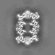
| Title | The cryoEM density map of T20s proteasome with various ice thickness subsets 4 and 5 combined (EMPIAR-10025 reprocessing) | |||||||||||||||
 Map data Map data | Sharpened and masked map | |||||||||||||||
 Sample Sample |
| |||||||||||||||
| Biological species |   Thermoplasma acidophilum (acidophilic) Thermoplasma acidophilum (acidophilic) | |||||||||||||||
| Method | single particle reconstruction / cryo EM / Resolution: 2.87 Å | |||||||||||||||
 Authors Authors | Olek M / Zhang P / Cowtan K / Chaban Y / Webb D | |||||||||||||||
| Funding support | European Union, 4 items
| |||||||||||||||
 Citation Citation | Journal: Structure / Year: 2022 Title: IceBreaker: Software for high-resolution single-particle cryo-EM with non-uniform ice. Authors: Mateusz Olek / Kevin Cowtan / Donovan Webb / Yuriy Chaban / Peijun Zhang /  Abstract: Despite the abundance of available software tools, optimal particle selection is still a vital issue in single-particle cryoelectron microscopy (cryo-EM). Regardless of the method used, most pickers ...Despite the abundance of available software tools, optimal particle selection is still a vital issue in single-particle cryoelectron microscopy (cryo-EM). Regardless of the method used, most pickers struggle when ice thickness varies on a micrograph. IceBreaker allows users to estimate the relative ice gradient and flatten it by equalizing the local contrast. It allows the differentiation of particles from the background and improves overall particle picking performance. Furthermore, we introduce an additional parameter corresponding to local ice thickness for each particle. Particles with a defined ice thickness can be grouped and filtered based on this parameter during processing. These functionalities are especially valuable for on-the-fly processing to automatically pick as many particles as possible from each micrograph and to select optimal regions for data collection. Finally, estimated ice gradient distributions can be stored separately and used to inspect the quality of prepared samples. #1: Journal: Elife / Year: 2015Title: 2.8 A resolution reconstruction of the Thermoplasma acidophilum 20S proteasome using cryo-electron microscopy. Authors: Campbell MG / Veesler D / Cheng A / Potter CS / Carragher B | |||||||||||||||
| History |
|
- Structure visualization
| Movie |
 Movie viewer Movie viewer |
|---|---|
| Structure viewer | EM map: SurfViewMolmilJmol/JSmol |
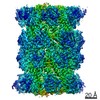
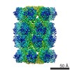
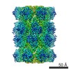
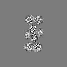
| Supplemental images |
- Downloads & links
-EMDB archive
| Map data | emd_13902.map.gz | 5.1 MB | EMDB map data format | |
|---|---|---|---|---|
| Header (meta data) | emd-13902-v30.xmlemd-13902.xml | 19.5 KB 19.5 KB | Display Display | EMDB header |
| FSC (resolution estimation) | emd_13902_fsc.xml | 7.7 KB | Display | FSC data file |
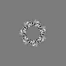
| Images |  emd_13902.png emd_13902.png | 166.6 KB | ||
| Masks | emd_13902_msk_1.map | 38.4 MB | Mask map | |
| Others | emd_13902_additional_1.map.gzemd_13902_additional_2.map.gzemd_13902_half_map_1.map.gzemd_13902_half_map_2.map.gz | 36 MB 29.5 MB 29.6 MB 29.5 MB | ||
| Archive directory |  http://ftp.pdbj.org/pub/emdb/structures/EMD-13902ftp://ftp.pdbj.org/pub/emdb/structures/EMD-13902 http://ftp.pdbj.org/pub/emdb/structures/EMD-13902ftp://ftp.pdbj.org/pub/emdb/structures/EMD-13902 | HTTPS FTP |
-Related structure data
| Related structure data | C: citing same article ( |
|---|---|
| Similar structure data |


-Links
| EMDB pages | EMDB (EBI/PDBe) / EMDataResource |
|---|
-Map
| File | Download / File: emd_13902.map.gz / Format: CCP4 / Size: 38.4 MB / Type: IMAGE STORED AS FLOATING POINT NUMBER (4 BYTES) | ||||||||||||||||||||||||||||||||||||||||||||||||||||||||||||
|---|---|---|---|---|---|---|---|---|---|---|---|---|---|---|---|---|---|---|---|---|---|---|---|---|---|---|---|---|---|---|---|---|---|---|---|---|---|---|---|---|---|---|---|---|---|---|---|---|---|---|---|---|---|---|---|---|---|---|---|---|---|
| Annotation | Sharpened and masked map | ||||||||||||||||||||||||||||||||||||||||||||||||||||||||||||
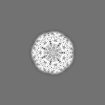
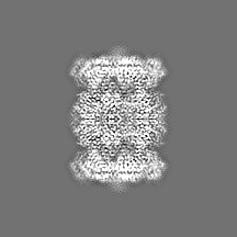
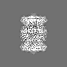
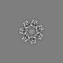
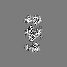
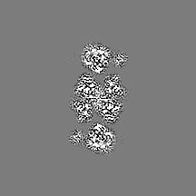
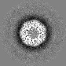
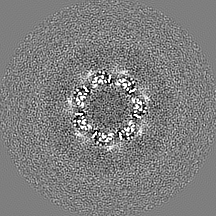
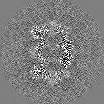
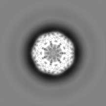
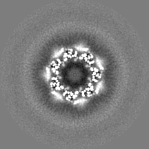
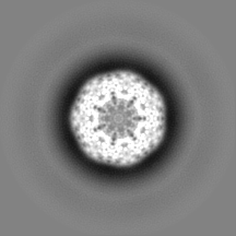
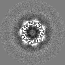
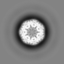
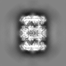
| Projections & slices | Image control
Images are generated by Spider. | ||||||||||||||||||||||||||||||||||||||||||||||||||||||||||||
| Voxel size | X=Y=Z: 1.315 Å | ||||||||||||||||||||||||||||||||||||||||||||||||||||||||||||
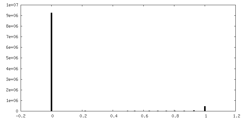
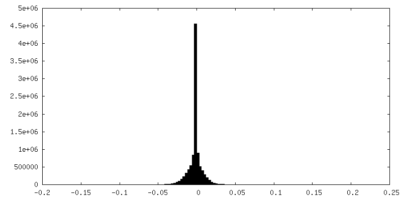
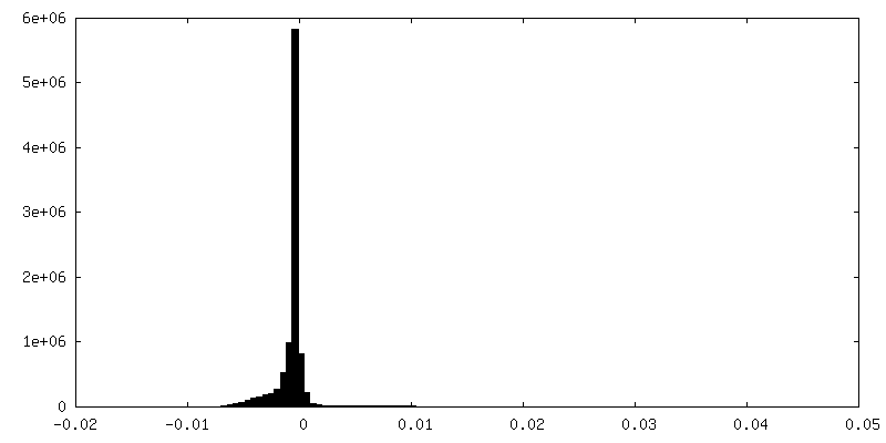
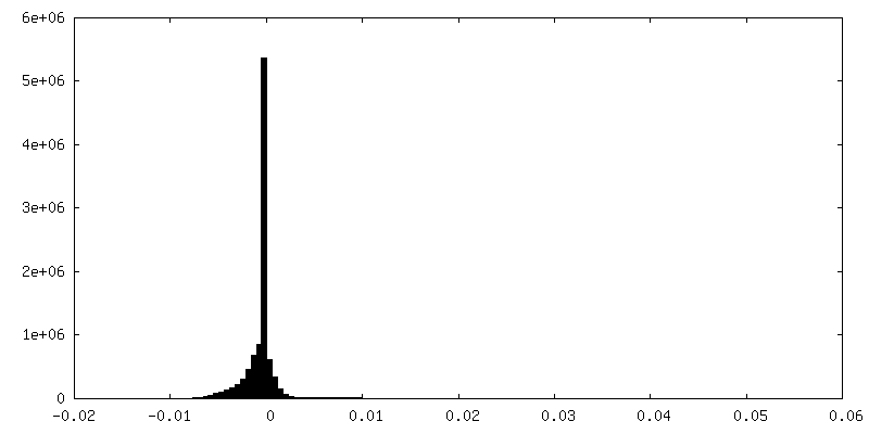
| Density |
| ||||||||||||||||||||||||||||||||||||||||||||||||||||||||||||
| Symmetry | Space group: 1 | ||||||||||||||||||||||||||||||||||||||||||||||||||||||||||||
| Details | EMDB XML:
CCP4 map header:
| ||||||||||||||||||||||||||||||||||||||||||||||||||||||||||||
 Z (Sec.)
 Y (Row.)
 X (Col.)


-Supplemental data
-Mask #1
| File | emd_13902_msk_1.map | ||||||||||||
|---|---|---|---|---|---|---|---|---|---|---|---|---|---|
| Projections & Slices |
| ||||||||||||
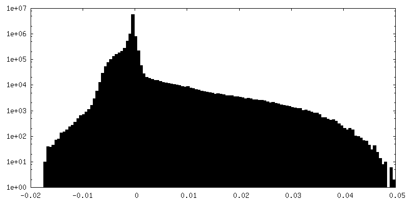
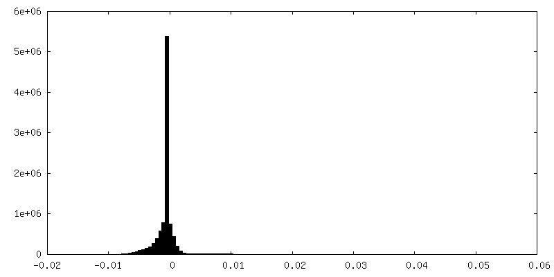
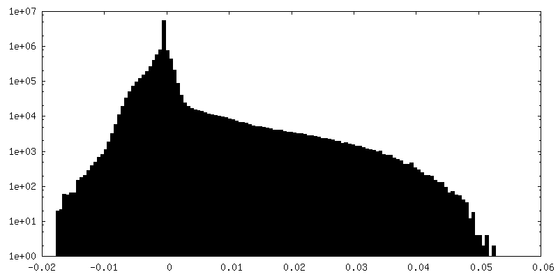
| Density Histograms |


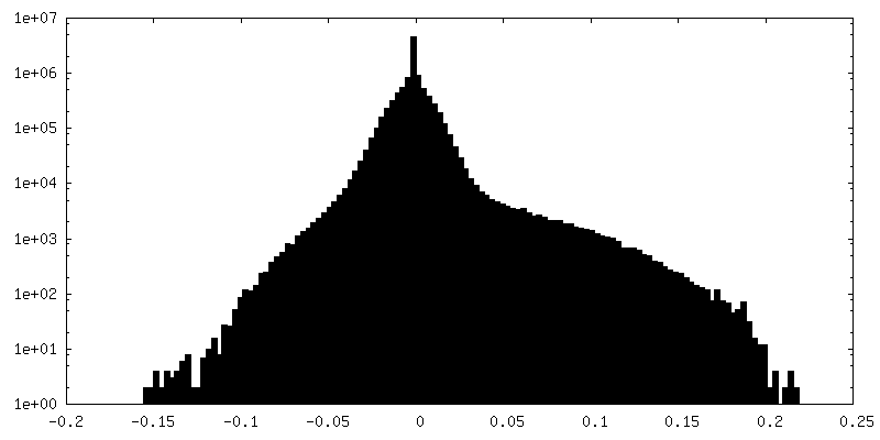
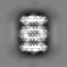
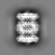
-Additional map: Sharpened map (not masked)
| File | emd_13902_additional_1.map | ||||||||||||
|---|---|---|---|---|---|---|---|---|---|---|---|---|---|
| Annotation | Sharpened map (not masked) | ||||||||||||
| Projections & Slices |
| ||||||||||||
| Density Histograms |
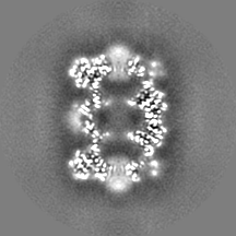
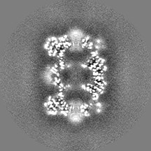
-Additional map: Unsharpened map
| File | emd_13902_additional_2.map | ||||||||||||
|---|---|---|---|---|---|---|---|---|---|---|---|---|---|
| Annotation | Unsharpened map | ||||||||||||
| Projections & Slices |
| ||||||||||||
| Density Histograms |
-Half map: Half map 1
| File | emd_13902_half_map_1.map | ||||||||||||
|---|---|---|---|---|---|---|---|---|---|---|---|---|---|
| Annotation | Half map 1 | ||||||||||||
| Projections & Slices |
| ||||||||||||
| Density Histograms |

-Half map: Half map 2
| File | emd_13902_half_map_2.map | ||||||||||||
|---|---|---|---|---|---|---|---|---|---|---|---|---|---|
| Annotation | Half map 2 | ||||||||||||
| Projections & Slices |
| ||||||||||||
| Density Histograms |
- Sample components
-Entire : Thermoplasma acidophilum 20S proteasome
| Entire | Name: Thermoplasma acidophilum 20S proteasome |
|---|---|
| Components |
|
-Supramolecule #1: Thermoplasma acidophilum 20S proteasome
| Supramolecule | Name: Thermoplasma acidophilum 20S proteasome / type: complex / ID: 1 / Parent: 0 |
|---|---|
| Source (natural) | Organism: Thermoplasma acidophilum (acidophilic) |
| Recombinant expression | Organism:  |
-Experimental details
-Structure determination
| Method | cryo EM |
|---|---|
 Processing Processing | single particle reconstruction |
| Aggregation state | particle |
-Sample preparation
| Concentration | 0.21 mg/mL |
|---|---|
| Buffer | pH: 7.8 / Details: 20 mM Tris, 150 mM NaCl |
| Vitrification | Cryogen name: ETHANE / Instrument: GATAN CRYOPLUNGE 3 |
- Electron microscopy
| Microscope | FEI TITAN KRIOS |
|---|---|
| Image recording | Film or detector model: GATAN K2 SUMMIT (4k x 4k) / Average electron dose: 53.0 e/Å2 |
| Electron beam | Acceleration voltage: 300 kV / Electron source:  FIELD EMISSION GUN FIELD EMISSION GUN |
| Electron optics | Illumination mode: FLOOD BEAM / Imaging mode: BRIGHT FIELD / Cs: 2.7 mm |
| Sample stage | Specimen holder model: FEI TITAN KRIOS AUTOGRID HOLDER |
| Experimental equipment |  Model: Titan Krios / Image courtesy: FEI Company |
-Image processing
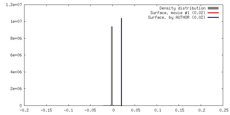
| Final reconstruction | Resolution.type: BY AUTHOR / Resolution: 2.87 Å / Resolution method: FSC 0.143 CUT-OFF / Number images used: 66651 |
|---|---|
| Initial angle assignment | Type: NOT APPLICABLE / Software - Name: RELION |
| Final angle assignment | Type: NOT APPLICABLE / Software - Name: RELION |
| FSC plot (resolution estimation) | 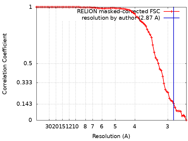 |
