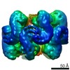
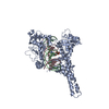
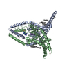
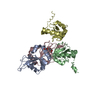
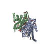
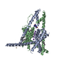
ジャーナル: Nature / 年: 2022 タイトル: Structure of the NLRP3 decamer bound to the cytokine release inhibitor CRID3. 著者: Inga V Hochheiser / Michael Pilsl / Gregor Hagelueken / Jonas Moecking / Michael Marleaux / Rebecca Brinkschulte / Eicke Latz / Christoph Engel / Matthias Geyer / 要旨: NLRP3 is an intracellular sensor protein that when activated by a broad spectrum of exogenous and endogenous stimuli leads to inflammasome formation and pyroptosis. The conformational states of NLRP3 ...NLRP3 is an intracellular sensor protein that when activated by a broad spectrum of exogenous and endogenous stimuli leads to inflammasome formation and pyroptosis. The conformational states of NLRP3 and the way antagonistic small molecules act at the molecular level remain poorly understood. Here we report the cryo-electron microscopy structures of full-length human NLRP3 in its native form and complexed with the inhibitor CRID3 (also named MCC950). Inactive, ADP-bound NLRP3 is a decamer composed of homodimers of intertwined leucine-rich repeat (LRR) domains that assemble back-to-back as pentamers. The NACHT domain is located at the apical axis of this spherical structure. One pyrin domain dimer is in addition formed inside the LRR cage. Molecular contacts between the concave sites of two opposing LRR domains are mediated by an acidic loop that extends from an LRR transition segment. Binding of CRID3 considerably stabilizes the NACHT and LRR domains relative to each other. CRID3 binds into a cleft, connecting four subdomains of the NACHT with the transition LRR. Its central sulfonylurea group interacts with the Walker A motif of the NLRP3 nucleotide-binding domain and is sandwiched between two arginine residues, which explains the specificity of NLRP3 for this chemical entity. With the determination of the binding site of this key therapeutic agent, specific targeting of NLRP3 for the treatment of autoinflammatory and autoimmune diseases and rational drug optimization is within reach.
UniProtKB: NACHT, LRR and PYD domains-containing protein 3
-
実験情報
-
構造解析
手法
クライオ電子顕微鏡法
解析
単粒子再構成法
試料の集合状態
particle
-
試料調製
濃度
0.01 mg/mL
緩衝液
pH: 7.5
凍結
凍結剤: ETHANE
詳細
purified protein
-
電子顕微鏡法
顕微鏡
FEI TITAN KRIOS
撮影
フィルム・検出器のモデル: GATAN K3 (6k x 4k) / 平均電子線量: 45.0 e/Å2
電子線
加速電圧: 300 kV / 電子線源: FIELD EMISSION GUN
電子光学系
照射モード: FLOOD BEAM / 撮影モード: BRIGHT FIELD
実験機器
モデル: Titan Krios / 画像提供: FEI Company
+
画像解析
初期モデル
モデルのタイプ: INSILICO MODEL
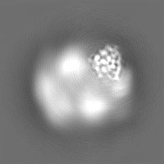
最終 再構成
解像度のタイプ: BY AUTHOR / 解像度: 4.1 Å / 解像度の算出法: FSC 0.143 CUT-OFF / ソフトウェア - 名称: RELION (ver. 3.1) 詳細: Focussed refinement of a NACHT domain of the human NLRP3 decamer 使用した粒子像数: 364354
初期 角度割当
タイプ: MAXIMUM LIKELIHOOD / ソフトウェア - 名称: RELION (ver. 3.1)
最終 角度割当
タイプ: MAXIMUM LIKELIHOOD / ソフトウェア - 名称: RELION (ver. 3.1)
 ムービー
ムービー コントローラー


 データを開く
 基本情報
基本情報 マップデータ
 試料
試料 キーワード
キーワード 機能・相同性情報
 Homo sapiens (ヒト)
 データ登録者
 ドイツ, 1件
  引用
引用 構造の表示
構造の表示
 ダウンロードとリンク
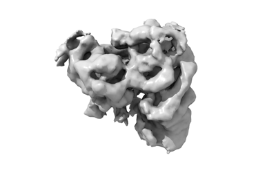 emd_13685.png
 http://ftp.pdbj.org/pub/emdb/structures/EMD-13685


 Z (Sec.)
 Y (Row.)
 X (Col.)


 試料の構成要素
 解析
解析 電子顕微鏡法
 FIELD EMISSION GUN

