 Movie
Movie Controller


+ Open data

- Basic information
| Entry | Database: PDB / ID: 6v3c | ||||||
|---|---|---|---|---|---|---|---|
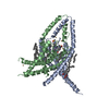
| Title | K2P2.1(TREK-1)I110D:Ru360 bound channel structure | ||||||
 Components Components | Potassium channel subfamily K member 2 | ||||||
 Keywords Keywords | MEMBRANE PROTEIN / K channel / TREK-1 / Mus musculus | ||||||
| Function / homology |  Function and homology information Function and homology informationTWIK related potassium channel (TREK) / Phase 4 - resting membrane potential / cellular response to arachidonate / mechanosensitive potassium channel activity / negative regulation of DNA biosynthetic process / detection of mechanical stimulus involved in sensory perception of touch / chemical synaptic transmission, postsynaptic / stabilization of membrane potential / cardiac ventricle development / ligand-gated channel activity ...TWIK related potassium channel (TREK) / Phase 4 - resting membrane potential / cellular response to arachidonate / mechanosensitive potassium channel activity / negative regulation of DNA biosynthetic process / detection of mechanical stimulus involved in sensory perception of touch / chemical synaptic transmission, postsynaptic / stabilization of membrane potential / cardiac ventricle development / ligand-gated channel activity / negative regulation of cardiac muscle cell proliferation / positive regulation of cellular response to hypoxia / potassium ion leak channel activity / glutamate secretion / node of Ranvier / outward rectifier potassium channel activity / potassium channel inhibitor activity / astrocyte projection / regulation of synaptic transmission, GABAergic / cochlea development / response to axon injury / voltage-gated potassium channel activity / neuronal action potential / voltage-gated potassium channel complex / potassium ion transmembrane transport / calyx of Held / axon terminus / chloride transmembrane transport / potassium ion transport / regulation of membrane potential / sarcolemma / postsynaptic density membrane / Schaffer collateral - CA1 synapse / memory / cellular response to hypoxia / apical plasma membrane / G protein-coupled receptor signaling pathway / protein heterodimerization activity / axon / neuronal cell body / dendrite / cell surface / endoplasmic reticulum / metal ion binding / identical protein binding / plasma membrane Similarity search - Function | ||||||
| Biological species |  | ||||||
| Method |  X-RAY DIFFRACTION / SYNCHROTRON / MOLECULAR REPLACEMENT / Resolution: 3.51 Å X-RAY DIFFRACTION / SYNCHROTRON / MOLECULAR REPLACEMENT / Resolution: 3.51 Å | ||||||
 Authors Authors | Pope, L. / Lolicato, M. / Minor, D.L. | ||||||
| Funding support |  United States, 1items United States, 1items
| ||||||
 Citation Citation | Journal: Cell Chem Biol / Year: 2020 Title: Polynuclear Ruthenium Amines Inhibit K2PChannels via a "Finger in the Dam" Mechanism. Authors: Pope, L. / Lolicato, M. / Minor Jr., D.L. | ||||||
| History |
|
- Structure visualization
| Structure viewer | Molecule: MolmilJmol/JSmol |
|---|
- Downloads & links
-Download
| PDBx/mmCIF format | 6v3c.cif.gz | 117.9 KB | Display | PDBx/mmCIF format |
|---|---|---|---|---|
| PDB format | pdb6v3c.ent.gz | 88.4 KB | Display | PDB format |
| PDBx/mmJSON format | 6v3c.json.gz | Tree view | PDBx/mmJSON format | |
| Others |  Other downloads Other downloads |
-Validation report
| Arichive directory | https://data.pdbj.org/pub/pdb/validation_reports/v3/6v3cftp://data.pdbj.org/pub/pdb/validation_reports/v3/6v3c | HTTPS FTP |
|---|
-Related structure data
| Related structure data |  6v36C  6v37C  6v3iC  4rueS S: Starting model for refinement C: citing same article ( |
|---|---|
| Similar structure data |


-Links
 PDBj
PDBj


- Assembly
| Deposited unit | 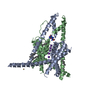
| ||||||||
|---|---|---|---|---|---|---|---|---|---|
| 1 |
| ||||||||
| Unit cell |
|
-Components
-Protein , 1 types, 2 molecules AB
| #1: Protein | Mass: 34305.867 Da / Num. of mol.: 2 Source method: isolated from a genetically manipulated source Source: (gene. exp.)  Komagataella pastoris (fungus) / References: UniProt: P97438*PLUS Komagataella pastoris (fungus) / References: UniProt: P97438*PLUS |
|---|
-Non-polymers , 7 types, 17 molecules 


| #2: Chemical | ChemComp-RU3 /  Mass: 436.355 Da / Num. of mol.: 1 / Source method: obtained synthetically / Formula: C2H18N8O5Ru2 / Feature type: SUBJECT OF INVESTIGATION Mass: 436.355 Da / Num. of mol.: 1 / Source method: obtained synthetically / Formula: C2H18N8O5Ru2 / Feature type: SUBJECT OF INVESTIGATION | ||||||||||
|---|---|---|---|---|---|---|---|---|---|---|---|
| #3: Chemical |  Mass: 112.411 Da / Num. of mol.: 3 / Source method: obtained synthetically / Formula: Cd Mass: 112.411 Da / Num. of mol.: 3 / Source method: obtained synthetically / Formula: Cd#4: Chemical | ChemComp-K /  Mass: 39.098 Da / Num. of mol.: 5 / Source method: obtained synthetically / Formula: K Mass: 39.098 Da / Num. of mol.: 5 / Source method: obtained synthetically / Formula: K#5: Chemical | ChemComp-OCT /  Mass: 114.229 Da / Num. of mol.: 4 / Source method: obtained synthetically / Formula: C8H18 Mass: 114.229 Da / Num. of mol.: 4 / Source method: obtained synthetically / Formula: C8H18#6: Chemical | ChemComp-LNK / |  Mass: 72.149 Da / Num. of mol.: 1 / Source method: obtained synthetically / Formula: C5H12 Mass: 72.149 Da / Num. of mol.: 1 / Source method: obtained synthetically / Formula: C5H12#7: Chemical | ChemComp-HEX / |  Mass: 86.175 Da / Num. of mol.: 1 / Source method: obtained synthetically / Formula: C6H14 Mass: 86.175 Da / Num. of mol.: 1 / Source method: obtained synthetically / Formula: C6H14#8: Water | ChemComp-HOH / | Mass: 18.015 Da / Num. of mol.: 2 / Source method: isolated from a natural source / Formula: H2O |
-Details
| Has ligand of interest | Y |
|---|---|
| Has protein modification | Y |
-Experimental details
-Experiment
| Experiment | Method: X-RAY DIFFRACTION / Number of used crystals: 1 |
|---|
- Sample preparation
| Crystal | Density Matthews: 3.79 Å3/Da / Density % sol: 67.56 % |
|---|---|
| Crystal grow | Temperature: 277 K / Method: vapor diffusion, hanging drop Details: 0.1 M KCl, 1-3 mM CdCl2, 0.1 M HEPES 7-8, 20-25% PEG400 |
-Data collection
| Diffraction | Mean temperature: 100 K / Serial crystal experiment: N |
|---|---|
| Diffraction source | Source: SYNCHROTRON / Site: APS / Beamline: 23-ID-B / Wavelength: 1.0332 Å |
| Detector | Type: DECTRIS EIGER X 16M / Detector: PIXEL / Date: Aug 18, 2018 |
| Radiation | Protocol: SINGLE WAVELENGTH / Monochromatic (M) / Laue (L): M / Scattering type: x-ray |
| Radiation wavelength | Wavelength: 1.0332 Å / Relative weight: 1 |
| Reflection | Resolution: 3.51→46.4 Å / Num. obs: 12595 / % possible obs: 100 % / Redundancy: 13.3 % / CC1/2: 1 / Net I/σ(I): 10.8 |
| Reflection shell | Resolution: 3.51→3.85 Å / Num. unique obs: 530 / CC1/2: 0.149 |
- Processing
| Software |
| |||||||||||||||||||||||||||||||||||
|---|---|---|---|---|---|---|---|---|---|---|---|---|---|---|---|---|---|---|---|---|---|---|---|---|---|---|---|---|---|---|---|---|---|---|---|---|
| Refinement | Method to determine structure: MOLECULAR REPLACEMENT Starting model: 4RUE Resolution: 3.51→14.98 Å / SU ML: 0.6 / Cross valid method: THROUGHOUT / σ(F): 1.33 / Phase error: 44.65 / Stereochemistry target values: ML
| |||||||||||||||||||||||||||||||||||
| Solvent computation | Shrinkage radii: 0.9 Å / VDW probe radii: 1.11 Å / Solvent model: FLAT BULK SOLVENT MODEL | |||||||||||||||||||||||||||||||||||
| Displacement parameters | Biso max: 327.88 Å2 / Biso mean: 203.5724 Å2 / Biso min: 119.59 Å2 | |||||||||||||||||||||||||||||||||||
| Refinement step | Cycle: final / Resolution: 3.51→14.98 Å
| |||||||||||||||||||||||||||||||||||
| LS refinement shell | Refine-ID: X-RAY DIFFRACTION / Rfactor Rfree error: 0 / Total num. of bins used: 4
|
