Movie
Movie Controller

[English] 日本語
 Yorodumi
- EMDB-13464: Structure of the 70S-EF-G-GDP ribosome complex with tRNAs in chim... -
+ Open data

- Basic information
| Entry | Database: EMDB / ID: EMD-13464 | |||||||||
|---|---|---|---|---|---|---|---|---|---|---|
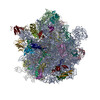
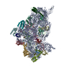
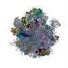
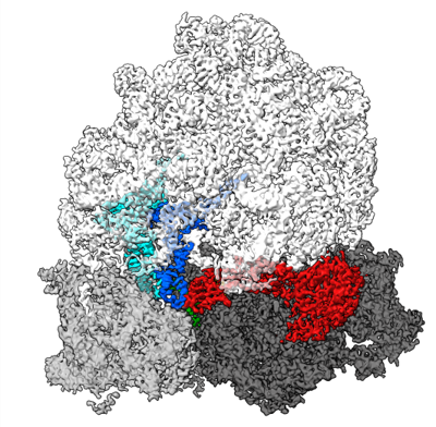
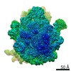
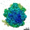
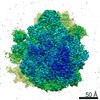
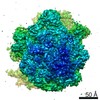
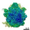
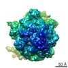
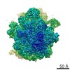
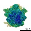
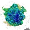
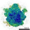
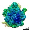
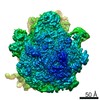
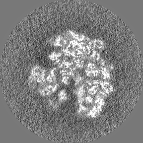
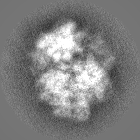
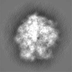
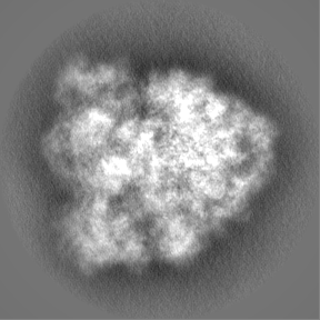
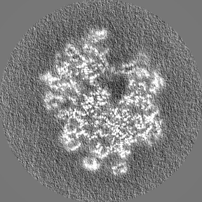
| Title | Structure of the 70S-EF-G-GDP ribosome complex with tRNAs in chimeric state 1 (CHI1-EF-G-GDP) | |||||||||
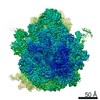
 Map data Map data | ||||||||||
 Sample Sample |
| |||||||||
 Keywords Keywords | EF-G / robosome / 70S / apramycin / translocation / RIBOSOME | |||||||||
| Function / homology |  Function and homology information Function and homology informationribosome disassembly / guanosine tetraphosphate binding / stringent response / negative regulation of cytoplasmic translational initiation / transcription antitermination factor activity, RNA binding / ornithine decarboxylase inhibitor activity / translational elongation / misfolded RNA binding / Group I intron splicing / RNA folding ...ribosome disassembly / guanosine tetraphosphate binding / stringent response / negative regulation of cytoplasmic translational initiation / transcription antitermination factor activity, RNA binding / ornithine decarboxylase inhibitor activity / translational elongation / misfolded RNA binding / Group I intron splicing / RNA folding / translation elongation factor activity / translational termination / transcriptional attenuation / endoribonuclease inhibitor activity / positive regulation of ribosome biogenesis / RNA-binding transcription regulator activity / four-way junction DNA binding / negative regulation of cytoplasmic translation / DnaA-L2 complex / regulation of mRNA stability / translation repressor activity / negative regulation of translational initiation / negative regulation of DNA-templated DNA replication initiation / mRNA regulatory element binding translation repressor activity / regulation of DNA-templated transcription elongation / positive regulation of RNA splicing / transcription elongation factor complex / response to reactive oxygen species / cytosolic ribosome assembly / ribosome assembly / assembly of large subunit precursor of preribosome / transcription antitermination / DNA endonuclease activity / translational initiation / regulation of cell growth / DNA-templated transcription termination / response to radiation / maintenance of translational fidelity / mRNA 5'-UTR binding / regulation of translation / large ribosomal subunit / transferase activity / ribosomal small subunit assembly / ribosome biogenesis / ribosome binding / ribosomal small subunit biogenesis / 5S rRNA binding / ribosomal large subunit assembly / small ribosomal subunit / small ribosomal subunit rRNA binding / cytosolic small ribosomal subunit / large ribosomal subunit rRNA binding / cytosolic large ribosomal subunit / Hydrolases; Acting on acid anhydrides; Acting on GTP to facilitate cellular and subcellular movement / cytoplasmic translation / tRNA binding / negative regulation of translation / rRNA binding / structural constituent of ribosome / ribosome / translation / response to antibiotic / negative regulation of DNA-templated transcription / hydrolase activity / mRNA binding / GTPase activity / GTP binding / DNA binding / RNA binding / zinc ion binding / membrane / cytoplasm / cytosol Similarity search - Function | |||||||||
| Biological species |  | |||||||||
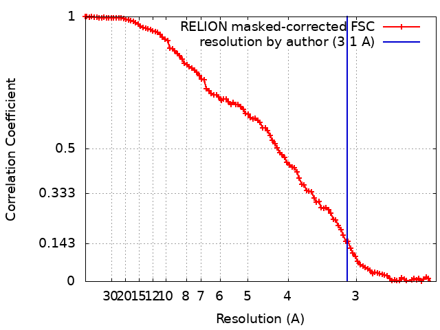
| Method | single particle reconstruction / cryo EM / Resolution: 3.1 Å | |||||||||
 Authors Authors | Petrychenko V / Peng BZ | |||||||||
| Funding support |  Germany, 2 items Germany, 2 items
| |||||||||
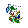
 Citation Citation | Journal: Nat Commun / Year: 2021 Title: Structural mechanism of GTPase-powered ribosome-tRNA movement. Authors: Valentyn Petrychenko / Bee-Zen Peng / Ana C de A P Schwarzer / Frank Peske / Marina V Rodnina / Niels Fischer / Abstract: GTPases are regulators of cell signaling acting as molecular switches. The translational GTPase EF-G stands out, as it uses GTP hydrolysis to generate force and promote the movement of the ribosome ...GTPases are regulators of cell signaling acting as molecular switches. The translational GTPase EF-G stands out, as it uses GTP hydrolysis to generate force and promote the movement of the ribosome along the mRNA. The key unresolved question is how GTP hydrolysis drives molecular movement. Here, we visualize the GTPase-powered step of ongoing translocation by time-resolved cryo-EM. EF-G in the active GDP-Pi form stabilizes the rotated conformation of ribosomal subunits and induces twisting of the sarcin-ricin loop of the 23 S rRNA. Refolding of the GTPase switch regions upon Pi release initiates a large-scale rigid-body rotation of EF-G pivoting around the sarcin-ricin loop that facilitates back rotation of the ribosomal subunits and forward swiveling of the head domain of the small subunit, ultimately driving tRNA forward movement. The findings demonstrate how a GTPase orchestrates spontaneous thermal fluctuations of a large RNA-protein complex into force-generating molecular movement. | |||||||||
| History |
|
- Structure visualization
| Movie |
Movie viewer |
|---|---|
| Structure viewer | EM map: SurfViewMolmilJmol/JSmol |
| Supplemental images |
- Downloads & links
-EMDB archive
| Map data | emd_13464.map.gz | 463.9 MB | EMDB map data format | |
|---|---|---|---|---|
| Header (meta data) | emd-13464-v30.xmlemd-13464.xml | 89.1 KB 89.1 KB | Display Display | EMDB header |
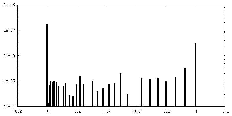
| FSC (resolution estimation) | emd_13464_fsc.xml | 10.2 KB | Display | FSC data file |
| Images |  emd_13464.png emd_13464.png | 204.8 KB | ||
| Masks | emd_13464_msk_1.map | 91.1 MB | Mask map | |
| Filedesc metadata | emd-13464.cif.gz | 17 KB | ||
| Others | emd_13464_half_map_1.map.gzemd_13464_half_map_2.map.gz | 71.1 MB 71.3 MB | ||
| Archive directory |  http://ftp.pdbj.org/pub/emdb/structures/EMD-13464ftp://ftp.pdbj.org/pub/emdb/structures/EMD-13464 http://ftp.pdbj.org/pub/emdb/structures/EMD-13464ftp://ftp.pdbj.org/pub/emdb/structures/EMD-13464 | HTTPS FTP |
-Related structure data
| Related structure data |  7pjyMC  7pjsC  7pjtC  7pjuC  7pjvC  7pjwC  7pjxC 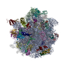 7pjzC M: atomic model generated by this map C: citing same article ( |
|---|---|
| Similar structure data | |
| EM raw data | EMPIAR-10792 (Title: Structural mechanism of GTPase-powered ribosome-tRNA movement Data size: 987.5 Data #1: Motion-corrected, dose-weighted micrographs [micrographs - single frame] Data #2: Shiny particles of non-rotated ribosome particles (C state), obtained by Bayesian polishing in Relion 3.1 [picked particles - single frame - processed] Data #3: Shiny particles of rotated ribosome particles, obtained by Bayesian polishing in Relion 3.1 [picked particles - single frame - processed]) |


-Links
| EMDB pages | EMDB (EBI/PDBe) / EMDataResource |
|---|---|
| Related items in Molecule of the Month |


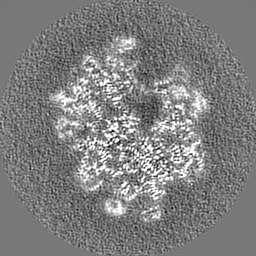
-Map
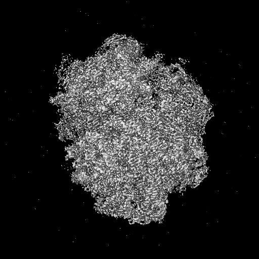
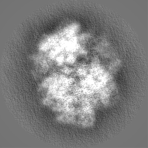
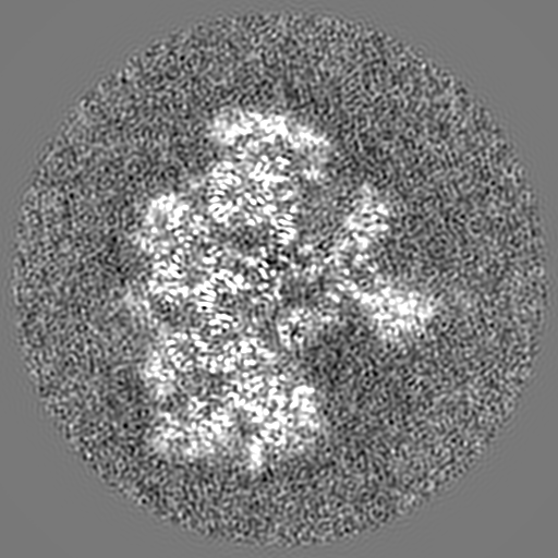
| File | Download / File: emd_13464.map.gz / Format: CCP4 / Size: 512 MB / Type: IMAGE STORED AS FLOATING POINT NUMBER (4 BYTES) | ||||||||||||||||||||||||||||||||||||||||||||||||||||||||||||||||||||
|---|---|---|---|---|---|---|---|---|---|---|---|---|---|---|---|---|---|---|---|---|---|---|---|---|---|---|---|---|---|---|---|---|---|---|---|---|---|---|---|---|---|---|---|---|---|---|---|---|---|---|---|---|---|---|---|---|---|---|---|---|---|---|---|---|---|---|---|---|---|
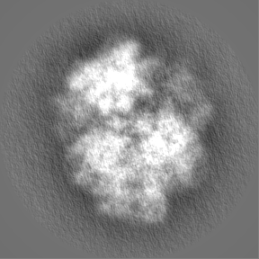
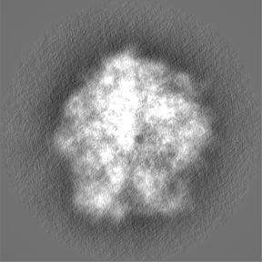
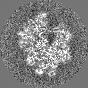
| Projections & slices | Image control
Images are generated by Spider. | ||||||||||||||||||||||||||||||||||||||||||||||||||||||||||||||||||||
| Voxel size | X=Y=Z: 0.6525 Å | ||||||||||||||||||||||||||||||||||||||||||||||||||||||||||||||||||||
| Density |
| ||||||||||||||||||||||||||||||||||||||||||||||||||||||||||||||||||||
| Symmetry | Space group: 1 | ||||||||||||||||||||||||||||||||||||||||||||||||||||||||||||||||||||
| Details | EMDB XML:
CCP4 map header:
| ||||||||||||||||||||||||||||||||||||||||||||||||||||||||||||||||||||
 Z (Sec.)
 Y (Row.)
 X (Col.)


-Supplemental data
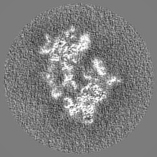
-Mask #1
| File | emd_13464_msk_1.map | ||||||||||||
|---|---|---|---|---|---|---|---|---|---|---|---|---|---|
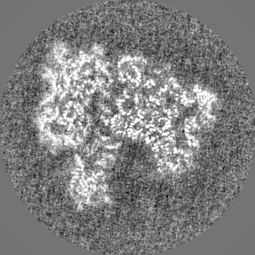
| Projections & Slices |
| ||||||||||||
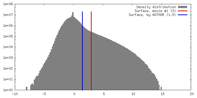
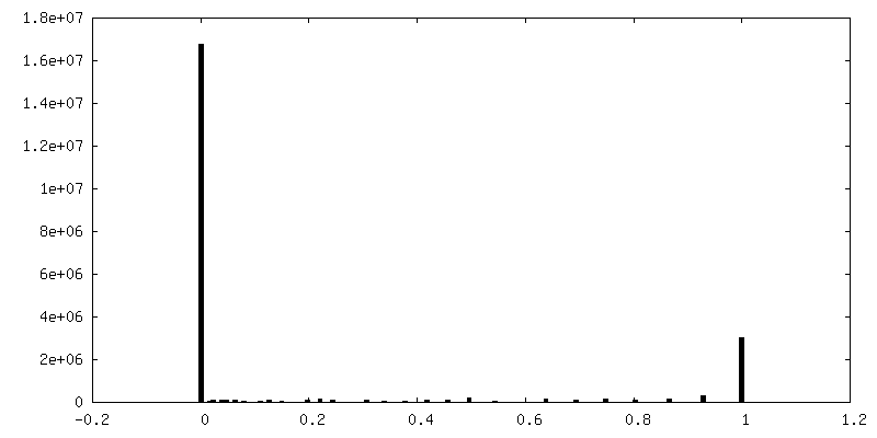
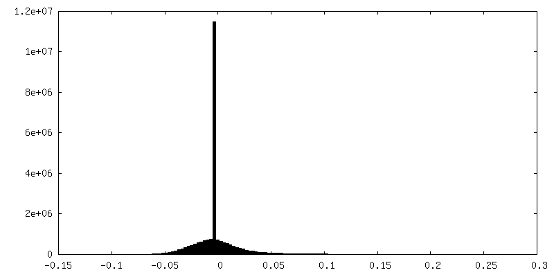
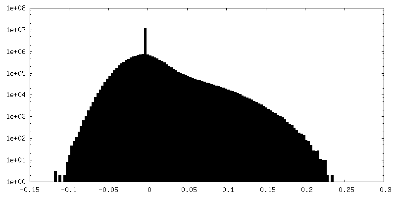
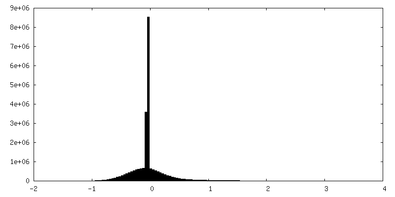
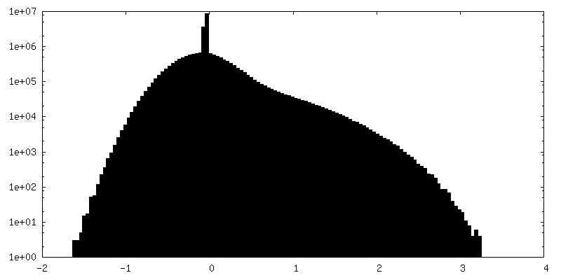
| Density Histograms |


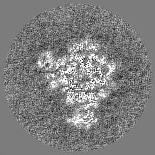
-Half map: #2
| File | emd_13464_half_map_1.map | ||||||||||||
|---|---|---|---|---|---|---|---|---|---|---|---|---|---|
| Projections & Slices |
| ||||||||||||
| Density Histograms |

-Half map: #1
| File | emd_13464_half_map_2.map | ||||||||||||
|---|---|---|---|---|---|---|---|---|---|---|---|---|---|
| Projections & Slices |
| ||||||||||||
| Density Histograms |


- Sample components
+Entire : E. coli ribosome complex 70S-Apr-EF-G-GDP-Pi-fMet-Phe-tRNAPhe-tRN...
+Supramolecule #1: E. coli ribosome complex 70S-Apr-EF-G-GDP-Pi-fMet-Phe-tRNAPhe-tRN...
+Macromolecule #1: 50S ribosomal protein L32
+Macromolecule #2: 50S ribosomal protein L33
+Macromolecule #3: 50S ribosomal protein L34
+Macromolecule #4: 50S ribosomal protein L35
+Macromolecule #5: 50S ribosomal protein L36
+Macromolecule #6: 50S ribosomal protein L10
+Macromolecule #7: 50S ribosomal protein L31
+Macromolecule #10: 50S ribosomal protein L2
+Macromolecule #11: 50S ribosomal protein L3
+Macromolecule #12: 50S ribosomal protein L4
+Macromolecule #13: 50S ribosomal protein L5
+Macromolecule #14: 50S ribosomal protein L6
+Macromolecule #15: 50S ribosomal protein L9
+Macromolecule #16: 50S ribosomal protein L11
+Macromolecule #17: 50S ribosomal protein L13
+Macromolecule #18: 50S ribosomal protein L14
+Macromolecule #19: 50S ribosomal protein L15
+Macromolecule #20: 50S ribosomal protein L16
+Macromolecule #21: 50S ribosomal protein L17
+Macromolecule #22: 50S ribosomal protein L18
+Macromolecule #23: 50S ribosomal protein L19
+Macromolecule #24: 50S ribosomal protein L20
+Macromolecule #25: 50S ribosomal protein L21
+Macromolecule #26: 50S ribosomal protein L22
+Macromolecule #27: 50S ribosomal protein L23
+Macromolecule #28: 50S ribosomal protein L24
+Macromolecule #29: 50S ribosomal protein L25
+Macromolecule #30: 50S ribosomal protein L27
+Macromolecule #31: 50S ribosomal protein L28
+Macromolecule #32: 50S ribosomal protein L29
+Macromolecule #33: 50S ribosomal protein L30
+Macromolecule #35: 30S ribosomal protein S2
+Macromolecule #36: 30S ribosomal protein S3
+Macromolecule #37: 30S ribosomal protein S4
+Macromolecule #38: 30S ribosomal protein S5
+Macromolecule #39: 30S ribosomal protein S6, fully modified isoform
+Macromolecule #40: 30S ribosomal protein S7
+Macromolecule #41: 30S ribosomal protein S8
+Macromolecule #42: 30S ribosomal protein S9
+Macromolecule #43: 30S ribosomal protein S10
+Macromolecule #44: 30S ribosomal protein S11
+Macromolecule #45: 30S ribosomal protein S12
+Macromolecule #46: 30S ribosomal protein S13
+Macromolecule #47: 30S ribosomal protein S14
+Macromolecule #48: 30S ribosomal protein S15
+Macromolecule #49: 30S ribosomal protein S16
+Macromolecule #50: 30S ribosomal protein S17
+Macromolecule #51: 30S ribosomal protein S18
+Macromolecule #52: 30S ribosomal protein S19
+Macromolecule #53: 30S ribosomal protein S20
+Macromolecule #54: 30S ribosomal protein S21
+Macromolecule #57: Elongation factor G
+Macromolecule #58: Dipeptide (FME-PHE)
+Macromolecule #8: 23S ribosomal RNA
+Macromolecule #9: 5S ribosomal RNA
+Macromolecule #34: 16S ribosomal RNA
+Macromolecule #55: P-site tRNA(fMet)
+Macromolecule #56: P-site fMet-Phe-tRNA(Phe)
+Macromolecule #59: mRNA
+Macromolecule #60: MAGNESIUM ION
+Macromolecule #61: ZINC ION
+Macromolecule #62: SODIUM ION
+Macromolecule #63: APRAMYCIN
+Macromolecule #64: GUANOSINE-5'-DIPHOSPHATE

-Experimental details
-Structure determination
| Method | cryo EM |
|---|---|
 Processing Processing | single particle reconstruction |
| Aggregation state | particle |
-Sample preparation
| Buffer | pH: 7.5 Details: 50 mM HEPES, 70 mM NH4Cl, 30 mM KCl, 3.5 mM MgCl2, 0.6 mM spermine, 0.4 mM spermidine |
|---|---|
| Grid | Model: Quantifoil R2/2 / Material: GOLD / Mesh: 200 / Pretreatment - Type: GLOW DISCHARGE / Pretreatment - Time: 15 sec. / Pretreatment - Atmosphere: OTHER |
| Vitrification | Cryogen name: ETHANE / Chamber humidity: 100 % / Chamber temperature: 277 K / Instrument: HOMEMADE PLUNGER / Details: Manual blotting & plunge-freezing. |
- Electron microscopy
| Microscope | FEI TITAN KRIOS |
|---|---|
| Details | Aberration corrections performed using Cs image corrector (CEOS company) |
| Image recording | Film or detector model: FEI FALCON III (4k x 4k) / Detector mode: INTEGRATING / Digitization - Dimensions - Width: 4096 pixel / Digitization - Dimensions - Height: 4096 pixel / Number grids imaged: 1 / Number real images: 8221 / Average exposure time: 1.0 sec. / Average electron dose: 30.0 e/Å2 |
| Electron beam | Acceleration voltage: 300 kV / Electron source:  FIELD EMISSION GUN FIELD EMISSION GUN |
| Electron optics | Calibrated defocus max: 1.2 µm / Calibrated defocus min: 0.5 µm / Illumination mode: FLOOD BEAM / Imaging mode: BRIGHT FIELD / Cs: 0.01 mm / Nominal defocus max: 1.5 µm / Nominal defocus min: 0.2 µm / Nominal magnification: 59000 |
| Sample stage | Specimen holder model: FEI TITAN KRIOS AUTOGRID HOLDER / Cooling holder cryogen: NITROGEN |
| Experimental equipment |  Model: Titan Krios / Image courtesy: FEI Company |
+Image processing
-Atomic model buiding 1
| Initial model |
| ||||||||||
|---|---|---|---|---|---|---|---|---|---|---|---|
| Details | Instead of Chimera ChimeraX was used. | ||||||||||
| Refinement | Space: REAL / Protocol: FLEXIBLE FIT / Target criteria: RSCC | ||||||||||
| Output model | PDB-7pjy: |