ムービー
ムービー コントローラー


+ データを開く

- 基本情報
基本情報
| 登録情報 | データベース: EMDB / ID: EMD-1340 | |||||||||
|---|---|---|---|---|---|---|---|---|---|---|
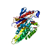
| タイトル | The beginning of kinesin's force-generating cycle visualized at 9-A resolution. | |||||||||
 マップデータ マップデータ | 9-Angstrom map of nucleotide-free kinesin complexed to the microtubule | |||||||||
 試料 試料 |
| |||||||||
| 機能・相同性 |  機能・相同性情報 機能・相同性情報regulation of modification of synapse structure, modulating synaptic transmission / plus-end-directed vesicle transport along microtubule / cytoplasm organization / cytolytic granule membrane / anterograde dendritic transport of neurotransmitter receptor complex / mitocytosis / anterograde axonal protein transport / anterograde neuronal dense core vesicle transport / retrograde neuronal dense core vesicle transport / ciliary rootlet ...regulation of modification of synapse structure, modulating synaptic transmission / plus-end-directed vesicle transport along microtubule / cytoplasm organization / cytolytic granule membrane / anterograde dendritic transport of neurotransmitter receptor complex / mitocytosis / anterograde axonal protein transport / anterograde neuronal dense core vesicle transport / retrograde neuronal dense core vesicle transport / ciliary rootlet / lysosome localization / positive regulation of potassium ion transport / plus-end-directed microtubule motor activity / RHO GTPases activate KTN1 / Kinesins / positive regulation of axon guidance / vesicle transport along microtubule / centrosome localization / mitochondrion transport along microtubule / microtubule motor activity / kinesin complex / COPI-dependent Golgi-to-ER retrograde traffic / natural killer cell mediated cytotoxicity / microtubule-based movement / stress granule disassembly / Insulin processing / synaptic vesicle transport / postsynaptic cytosol / microtubule-based process / phagocytic vesicle / axon cytoplasm / MHC class II antigen presentation / dendrite cytoplasm / axon guidance / positive regulation of synaptic transmission, GABAergic / positive regulation of protein localization to plasma membrane / sperm end piece / regulation of membrane potential / structural constituent of cytoskeleton / cellular response to type II interferon / microtubule cytoskeleton organization / neuron migration / centriolar satellite / Signaling by ALK fusions and activated point mutants / mitotic cell cycle / microtubule cytoskeleton / nuclear membrane / microtubule binding / vesicle / 加水分解酵素; 酸無水物に作用; GTPに作用・細胞または細胞小器官の運動に関与 / microtubule / cadherin binding / protein heterodimerization activity / hydrolase activity / GTPase activity / GTP binding / protein-containing complex binding / perinuclear region of cytoplasm / ATP hydrolysis activity / mitochondrion / ATP binding / membrane / metal ion binding / identical protein binding / cytoplasm / cytosol 類似検索 - 分子機能 | |||||||||
| 生物種 |  Homo sapiens (ヒト) Homo sapiens (ヒト) | |||||||||
| 手法 | 単粒子再構成法 / クライオ電子顕微鏡法 / 解像度: 9.0 Å | |||||||||
 データ登録者 データ登録者 | Sindelar CV / Downing KH | |||||||||
 引用 引用 | ジャーナル: J Cell Biol / 年: 2007 タイトル: The beginning of kinesin's force-generating cycle visualized at 9-A resolution. 著者: Charles V Sindelar / Kenneth H Downing /  要旨: We have used cryo-electron microscopy of kinesin-decorated microtubules to resolve the structure of the motor protein kinesin's crucial nucleotide response elements, switch I and the switch II helix, ...We have used cryo-electron microscopy of kinesin-decorated microtubules to resolve the structure of the motor protein kinesin's crucial nucleotide response elements, switch I and the switch II helix, in kinesin's poorly understood nucleotide-free state. Both of the switch elements undergo conformational change relative to the microtubule-free state. The changes in switch I suggest a role for it in "ejecting" adenosine diphosphate when kinesin initially binds to the microtubule. The switch II helix has an N-terminal extension, apparently stabilized by conserved microtubule contacts, implying a microtubule activation mechanism that could convey the state of the bound nucleotide to kinesin's putative force-delivering element (the "neck linker"). In deriving this structure, we have adapted an image-processing technique, single-particle reconstruction, for analyzing decorated microtubules. The resulting reconstruction visualizes the asymmetric seam present in native, 13-protofilament microtubules, and this method will provide an avenue to higher-resolution characterization of a variety of microtubule- binding proteins, as well as the microtubule itself. | |||||||||
| 履歴 |
|
- 構造の表示
構造の表示
| ムービー |
ムービービューア |
|---|---|
| 構造ビューア | EMマップ: SurfViewMolmilJmol/JSmol |
| 添付画像 |
 UCSF Chimera
- ダウンロードとリンク
-EMDBアーカイブ
| マップデータ | emd_1340.map.gz | 15.4 MB | EMDBマップデータ形式 | |
|---|---|---|---|---|
| ヘッダ (付随情報) | emd-1340-v30.xmlemd-1340.xml | 12.5 KB 12.5 KB | 表示 表示 | EMDBヘッダ |
| 画像 |  1340.gif 1340.gif | 90.8 KB | ||
| アーカイブディレクトリ |  http://ftp.pdbj.org/pub/emdb/structures/EMD-1340ftp://ftp.pdbj.org/pub/emdb/structures/EMD-1340 http://ftp.pdbj.org/pub/emdb/structures/EMD-1340ftp://ftp.pdbj.org/pub/emdb/structures/EMD-1340 | HTTPS FTP |
-関連構造データ


-リンク
| EMDBのページ | EMDB (EBI/PDBe) / EMDataResource |
|---|---|
| 「今月の分子」の関連する項目 |


-マップ
| ファイル | ダウンロード / ファイル: emd_1340.map.gz / 形式: CCP4 / 大きさ: 21.7 MB / タイプ: IMAGE STORED AS FLOATING POINT NUMBER (4 BYTES) | ||||||||||||||||||||||||||||||||||||||||||||||||||||||||||||||||||||
|---|---|---|---|---|---|---|---|---|---|---|---|---|---|---|---|---|---|---|---|---|---|---|---|---|---|---|---|---|---|---|---|---|---|---|---|---|---|---|---|---|---|---|---|---|---|---|---|---|---|---|---|---|---|---|---|---|---|---|---|---|---|---|---|---|---|---|---|---|---|
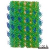
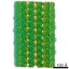
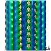
| 注釈 | 9-Angstrom map of nucleotide-free kinesin complexed to the microtubule | ||||||||||||||||||||||||||||||||||||||||||||||||||||||||||||||||||||
| 投影像・断面図 | 画像のコントロール
画像は Spider により作成 | ||||||||||||||||||||||||||||||||||||||||||||||||||||||||||||||||||||
| ボクセルのサイズ | X=Y=Z: 2 Å | ||||||||||||||||||||||||||||||||||||||||||||||||||||||||||||||||||||
| 密度 |
| ||||||||||||||||||||||||||||||||||||||||||||||||||||||||||||||||||||
| 対称性 | 空間群: 1 | ||||||||||||||||||||||||||||||||||||||||||||||||||||||||||||||||||||
| 詳細 | EMDB XML:
CCP4マップ ヘッダ情報:
| ||||||||||||||||||||||||||||||||||||||||||||||||||||||||||||||||||||
 Z (Sec.)
 Y (Row.)
 X (Col.)


-添付データ
- 試料の構成要素
-全体 : Nucleotide-free kinesin bound to a 13-protofilament microtubule
| 全体 | 名称: Nucleotide-free kinesin bound to a 13-protofilament microtubule |
|---|---|
| 要素 |
|
-超分子 #1000: Nucleotide-free kinesin bound to a 13-protofilament microtubule
| 超分子 | 名称: Nucleotide-free kinesin bound to a 13-protofilament microtubule タイプ: sample / ID: 1000 集合状態: The 13-protofilament microtubule forms a pseudo helix interrupted by a single seam Number unique components: 3 |
|---|
-分子 #1: Kinesin
| 分子 | 名称: Kinesin / タイプ: protein_or_peptide / ID: 1 / Name.synonym: K349 詳細: C220 "cys-lite" K349 construct: 6 of 8 native cysteines eliminated, introduced cysteine at position 220 コピー数: 1 / 集合状態: Monomer / 組換発現: Yes |
|---|---|
| 由来(天然) | 生物種: Homo sapiens (ヒト) / 別称: Human |
| 分子量 | 実験値: 36.4134 KDa |
| 組換発現 | 生物種:  |
-分子 #2: Alpha-tubulin
| 分子 | 名称: Alpha-tubulin / タイプ: protein_or_peptide / ID: 2 / 詳細: Glycerol-free tubulin purchased from Cytoskeleton / コピー数: 1 / 集合状態: Dimer / 組換発現: No / データベース: NCBI |
|---|---|
| 由来(天然) | 生物種: Homo sapiens (ヒト) / 別称: Human / 組織: Brain / 細胞中の位置: Brain |
| 分子量 | 実験値: 50.1279 KDa |
-分子 #3: Beta-tubulin
| 分子 | 名称: Beta-tubulin / タイプ: protein_or_peptide / ID: 3 / 詳細: Glycerol-free tubulin purchased from Cytoskeleton / コピー数: 1 / 集合状態: Dimer / 組換発現: No / データベース: NCBI |
|---|---|
| 由来(天然) | 生物種: Homo sapiens (ヒト) / 別称: Human / 組織: Brain |
| 分子量 | 実験値: 49.9283 KDa |
-実験情報
-構造解析
| 手法 | クライオ電子顕微鏡法 |
|---|---|
 解析 解析 | 単粒子再構成法 |
| 試料の集合状態 | particle |
-試料調製
| 緩衝液 | pH: 6.8 / 詳細: 25mM Pipes, 25mM NaCl, 2mM MgCl2, 1mM EGTA |
|---|---|
| グリッド | 詳細: 300 mesh copper grid |
| 凍結 | 凍結剤: ETHANE / チャンバー内温度: 93 K / 装置: HOMEMADE PLUNGER / 詳細: Vitrification instrument: Home-built 手法: Grids were not glow discharged. Liquid was almost entirely wicked away prior to blotting. Blotting time was less than 0.5 sec. |
- 電子顕微鏡法
| 顕微鏡 | JEOL 4000EX |
|---|---|
| 温度 | 平均: 105 K |
| アライメント法 | Legacy - 非点収差: e.g objective lens astigmatism was corrected at 400,000 times magnification |
| 撮影 | カテゴリ: FILM / フィルム・検出器のモデル: KODAK SO-163 FILM / デジタル化 - スキャナー: OTHER / デジタル化 - サンプリング間隔: 6.3 µm / 実像数: 350 / 平均電子線量: 16 e/Å2 / ビット/ピクセル: 16 |
| Tilt angle min | 0 |
| Tilt angle max | 0 |
| 電子線 | 加速電圧: 400 kV / 電子線源: LAB6 |
| 電子光学系 | 照射モード: FLOOD BEAM / 撮影モード: BRIGHT FIELD / Cs: 4.1 mm / 最大 デフォーカス(公称値): 1.5 µm / 最小 デフォーカス(公称値): 0.8 µm / 倍率(公称値): 60000 |
| 試料ステージ | 試料ホルダー: Eucentric / 試料ホルダーモデル: GATAN LIQUID NITROGEN |
-画像解析
| CTF補正 | 詳細: CTF correction was integrated into the back-projection process |
|---|---|
| 最終 再構成 | アルゴリズム: OTHER / 解像度のタイプ: BY AUTHOR / 解像度: 9.0 Å / 解像度の算出法: FSC 0.5 CUT-OFF / ソフトウェア - 名称: SPIDER 詳細: Data from 188 microtubules were incorporated into the final reconstruction 使用した粒子像数: 150000 |
-原子モデル構築 1
| 初期モデル | (PDB ID: , ) |
|---|---|
| ソフトウェア | 名称: SITUS |
| 詳細 | Protocol: Rigid body. Kinesin and tubulin were separately fitted into the final map using the program SITUS. |
| 精密化 | 空間: REAL / プロトコル: RIGID BODY FIT / 当てはまり具合の基準: Cross-correlation |
| 得られたモデル | 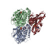 PDB-2p4n: |