Biotechnology and Biological Sciences Research Council (BBSRC)
United Kingdom
Citation
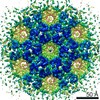
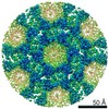
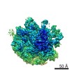
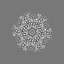
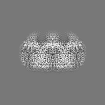
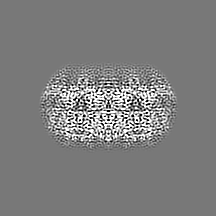
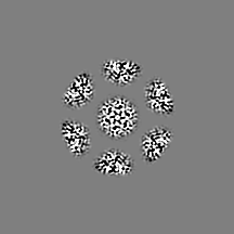
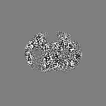
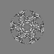
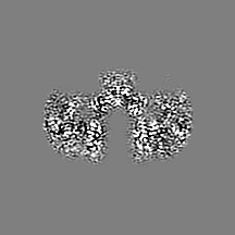
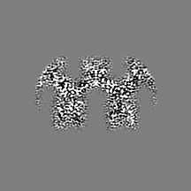
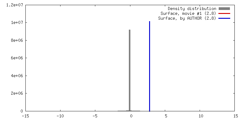
Journal: Nat Protoc / Year: 2022 Title: High-resolution in situ structure determination by cryo-electron tomography and subtomogram averaging using emClarity. Authors: Tao Ni / Thomas Frosio / Luiza Mendonça / Yuewen Sheng / Daniel Clare / Benjamin A Himes / Peijun Zhang / Abstract: Cryo-electron tomography and subtomogram averaging (STA) has developed rapidly in recent years. It provides structures of macromolecular complexes in situ and in cellular context at or below ...Cryo-electron tomography and subtomogram averaging (STA) has developed rapidly in recent years. It provides structures of macromolecular complexes in situ and in cellular context at or below subnanometer resolution and has led to unprecedented insights into the inner working of molecular machines in their native environment, as well as their functional relevant conformations and spatial distribution within biological cells or tissues. Given the tremendous potential of cryo-electron tomography STA in in situ structural cell biology, we previously developed emClarity, a graphics processing unit-accelerated image-processing software that offers STA and classification of macromolecular complexes at high resolution. However, the workflow remains challenging, especially for newcomers to the field. In this protocol, we describe a detailed workflow, processing and parameters associated with each step, from initial tomography tilt-series data to the final 3D density map, with several features unique to emClarity. We use four different samples, including human immunodeficiency virus type 1 Gag assemblies, ribosome and apoferritin, to illustrate the procedure and results of STA and classification. Following the processing steps described in this protocol, along with a comprehensive tutorial and guidelines for troubleshooting and parameter optimization, one can obtain density maps up to 2.8 Å resolution from six tilt series by cryo-electron tomography STA.
History
Deposition
Aug 10, 2021
-
Header (metadata) release
Feb 2, 2022
-
Map release
Feb 2, 2022
-
Update
Feb 23, 2022
-
Current status
Feb 23, 2022
Processing site: PDBe / Status: Released
-
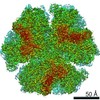
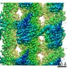
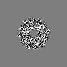
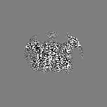
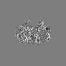
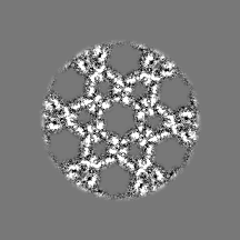
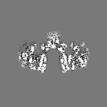
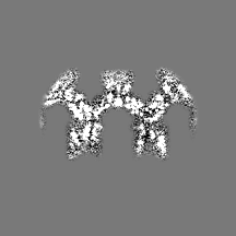
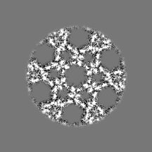
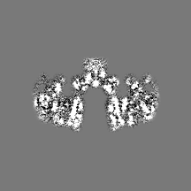
Structure visualization
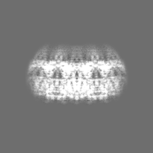
Movie
Surface view with section colored by density value
In the structure databanks used in Yorodumi, some data are registered as the other names, "COVID-19 virus" and "2019-nCoV". Here are the details of the virus and the list of structure data.
Jan 31, 2019. EMDB accession codes are about to change! (news from PDBe EMDB page)
EMDB accession codes are about to change! (news from PDBe EMDB page)
The allocation of 4 digits for EMDB accession codes will soon come to an end. Whilst these codes will remain in use, new EMDB accession codes will include an additional digit and will expand incrementally as the available range of codes is exhausted. The current 4-digit format prefixed with “EMD-” (i.e. EMD-XXXX) will advance to a 5-digit format (i.e. EMD-XXXXX), and so on. It is currently estimated that the 4-digit codes will be depleted around Spring 2019, at which point the 5-digit format will come into force.
The EM Navigator/Yorodumi systems omit the EMD- prefix.
Related info.:Q: What is EMD? / ID/Accession-code notation in Yorodumi/EM Navigator
Yorodumi is a browser for structure data from EMDB, PDB, SASBDB, etc.
This page is also the successor to EM Navigator detail page, and also detail information page/front-end page for Omokage search.
The word "yorodu" (or yorozu) is an old Japanese word meaning "ten thousand". "mi" (miru) is to see.
Related info.:EMDB / PDB / SASBDB / Comparison of 3 databanks / Yorodumi Search / Aug 31, 2016. New EM Navigator & Yorodumi / Yorodumi Papers / Jmol/JSmol / Function and homology information / Changes in new EM Navigator and Yorodumi
 Movie
Movie Controller

 Yorodumi
 Open data
 Basic information
 Map data
 Sample
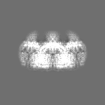
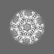
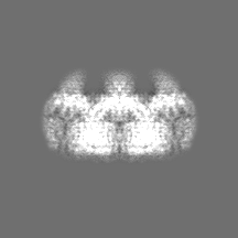
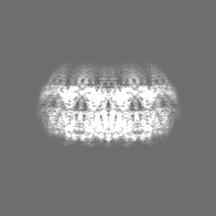
 Human immunodeficiency virus 1
 Authors
 United Kingdom, 2 items
  Citation
 Structure visualization
 Movie viewer
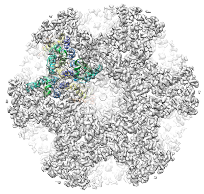
 Downloads & links
 emd_13354.png
 http://ftp.pdbj.org/pub/emdb/structures/EMD-13354
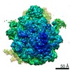


 Z (Sec.)
 Y (Row.)
 X (Col.)


 Sample components
 Processing
 Electron microscopy
 FIELD EMISSION GUN