Journal: EMBO J / Year: 2021 Title: Neutralization of SARS-CoV-2 by highly potent, hyperthermostable, and mutation-tolerant nanobodies. Authors: Thomas Güttler / Metin Aksu / Antje Dickmanns / Kim M Stegmann / Kathrin Gregor / Renate Rees / Waltraud Taxer / Oleh Rymarenko / Jürgen Schünemann / Christian Dienemann / Philip Gunkel / ...Authors: Thomas Güttler / Metin Aksu / Antje Dickmanns / Kim M Stegmann / Kathrin Gregor / Renate Rees / Waltraud Taxer / Oleh Rymarenko / Jürgen Schünemann / Christian Dienemann / Philip Gunkel / Bianka Mussil / Jens Krull / Ulrike Teichmann / Uwe Groß / Volker C Cordes / Matthias Dobbelstein / Dirk Görlich / Abstract: Monoclonal anti-SARS-CoV-2 immunoglobulins represent a treatment option for COVID-19. However, their production in mammalian cells is not scalable to meet the global demand. Single-domain (VHH) ...Monoclonal anti-SARS-CoV-2 immunoglobulins represent a treatment option for COVID-19. However, their production in mammalian cells is not scalable to meet the global demand. Single-domain (VHH) antibodies (also called nanobodies) provide an alternative suitable for microbial production. Using alpaca immune libraries against the receptor-binding domain (RBD) of the SARS-CoV-2 Spike protein, we isolated 45 infection-blocking VHH antibodies. These include nanobodies that can withstand 95°C. The most effective VHH antibody neutralizes SARS-CoV-2 at 17-50 pM concentration (0.2-0.7 µg per liter), binds the open and closed states of the Spike, and shows a tight RBD interaction in the X-ray and cryo-EM structures. The best VHH trimers neutralize even at 40 ng per liter. We constructed nanobody tandems and identified nanobody monomers that tolerate the K417N/T, E484K, N501Y, and L452R immune-escape mutations found in the Alpha, Beta, Gamma, Epsilon, Iota, and Delta/Kappa lineages. We also demonstrate neutralization of the Beta strain at low-picomolar VHH concentrations. We further discovered VHH antibodies that enforce native folding of the RBD in the E. coli cytosol, where its folding normally fails. Such "fold-promoting" nanobodies may allow for simplified production of vaccines and their adaptation to viral escape-mutations.
History
Deposition
Jun 23, 2021
-
Header (metadata) release
Aug 11, 2021
-
Map release
Aug 11, 2021
-
Update
Oct 13, 2021
-
Current status
Oct 13, 2021
Processing site: PDBe / Status: Released
-
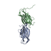
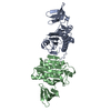
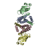
Structure visualization
Movie
Surface view with section colored by density value
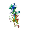
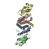
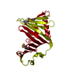
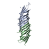
Macromolecule #1: SARS-CoV-2 HexaPro S (Spike) glycoprotein
Macromolecule
Name: SARS-CoV-2 HexaPro S (Spike) glycoprotein / type: protein_or_peptide / ID: 1 Details: Spike ectodomain (1-1208) with six exchanges to proline (F817P, A892P, A899P, A942P, K986P, V986P), a GSAS substitution at the furin cleavage site (residues 682-685), a C-terminal T4 foldon ...Details: Spike ectodomain (1-1208) with six exchanges to proline (F817P, A892P, A899P, A942P, K986P, V986P), a GSAS substitution at the furin cleavage site (residues 682-685), a C-terminal T4 foldon trimerization domain, followed by a HRV 3C protease site, a His8-tag and a Twin-Strep tag Enantiomer: LEVO
Source (natural)
Organism: Severe acute respiratory syndrome coronavirus 2
Cryogen name: ETHANE / Chamber humidity: 100 % / Chamber temperature: 277.15 K / Instrument: FEI VITROBOT MARK IV Details: sample volume: 3.0 microliters blotting time: 4 s blot force setting: 5.
Details
The Spike protein was mixed with a 9-fold molar excess of VHH Re5D06 and purified by size exclusion chromatography (Superose 6 Increase 3.2/300, Cytiva). The peak eluate fraction (1 mg/ml) was immediately applied to a freshly glow-discharged grid.
-
Electron microscopy
Microscope
FEI TITAN KRIOS
Specialist optics
Energy filter - Name: GIF Quantum LS / Energy filter - Slit width: 20 eV
Image recording
Film or detector model: GATAN K3 (6k x 4k) / Digitization - Dimensions - Width: 5760 pixel / Digitization - Dimensions - Height: 4092 pixel / Digitization - Sampling interval: 5.0 µm / Number grids imaged: 1 / Number real images: 15636 / Average exposure time: 2.0 sec. / Average electron dose: 39.91 e/Å2 Details: counting mode (non-super-resolution) 4 images per hole (beam-image shift)
Electron beam
Acceleration voltage: 300 kV / Electron source: FIELD EMISSION GUN
Type of model: OTHER Details: An ab-initio 3D model was generated from particles of good 2D classes using cryoSPARC (version 2.15).
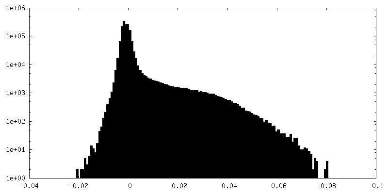
Final reconstruction
Number classes used: 3 / Applied symmetry - Point group: C1 (asymmetric) / Algorithm: FOURIER SPACE / Resolution.type: BY AUTHOR / Resolution: 3.5 Å / Resolution method: FSC 0.143 CUT-OFF / Software - Name: RELION (ver. 3.1) / Number images used: 652015
Initial angle assignment
Type: MAXIMUM LIKELIHOOD / Software - Name: cryoSPARC (ver. 2.15)
Final angle assignment
Type: MAXIMUM LIKELIHOOD / Software - Name: RELION (ver. 3.1)
Final 3D classification
Number classes: 5 / Software - Name: RELION (ver. 3.1) Details: The RBD-nanobody sub-volume (for the RBD-VHH Re5D06 module in the down conformation) was masked for focused 3D classification, which yielded 5 classes. Three classes were selected for ...Details: The RBD-nanobody sub-volume (for the RBD-VHH Re5D06 module in the down conformation) was masked for focused 3D classification, which yielded 5 classes. Three classes were selected for subsequent global and multi-body refinement runs.
FSC plot (resolution estimation)
+
About Yorodumi
-
News
-
Feb 9, 2022. New format data for meta-information of EMDB entries
New format data for meta-information of EMDB entries
Version 3 of the EMDB header file is now the official format.
The previous official version 1.9 will be removed from the archive.
In the structure databanks used in Yorodumi, some data are registered as the other names, "COVID-19 virus" and "2019-nCoV". Here are the details of the virus and the list of structure data.
Jan 31, 2019. EMDB accession codes are about to change! (news from PDBe EMDB page)
EMDB accession codes are about to change! (news from PDBe EMDB page)
The allocation of 4 digits for EMDB accession codes will soon come to an end. Whilst these codes will remain in use, new EMDB accession codes will include an additional digit and will expand incrementally as the available range of codes is exhausted. The current 4-digit format prefixed with “EMD-” (i.e. EMD-XXXX) will advance to a 5-digit format (i.e. EMD-XXXXX), and so on. It is currently estimated that the 4-digit codes will be depleted around Spring 2019, at which point the 5-digit format will come into force.
The EM Navigator/Yorodumi systems omit the EMD- prefix.
Related info.:Q: What is EMD? / ID/Accession-code notation in Yorodumi/EM Navigator
Yorodumi is a browser for structure data from EMDB, PDB, SASBDB, etc.
This page is also the successor to EM Navigator detail page, and also detail information page/front-end page for Omokage search.
The word "yorodu" (or yorozu) is an old Japanese word meaning "ten thousand". "mi" (miru) is to see.
Related info.:EMDB / PDB / SASBDB / Comparison of 3 databanks / Yorodumi Search / Aug 31, 2016. New EM Navigator & Yorodumi / Yorodumi Papers / Jmol/JSmol / Function and homology information / Changes in new EM Navigator and Yorodumi
 Movie
Movie Controller

 Yorodumi
 Open data
 Basic information
 Map data
 Sample
 Function and homology information
 Severe acute respiratory syndrome coronavirus 2 /
 Authors
 Germany, 1 items
  Citation
 Structure visualization
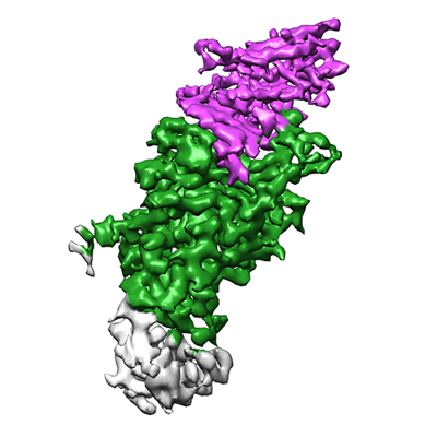
 Downloads & links
 emd_13105.png
 http://ftp.pdbj.org/pub/emdb/structures/EMD-13105


 Z (Sec.)
 Y (Row.)
 X (Col.)


 Sample components
 Homo sapiens (human) / Recombinant strain: Expi293F / Recombinant plasmid: Addgene #154745
 Komagataella pastoris (fungus)
 Processing
 Electron microscopy
 FIELD EMISSION GUN

