 Movie
Movie Controller

[English] 日本語
 Yorodumi
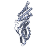
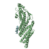
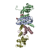
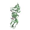
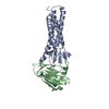
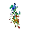
- PDB-5vtg: The structure of TamB963-1138 from Escherichia coli reveals a nov... -
+ Open data

- Basic information
| Entry | Database: PDB / ID: 5vtg | ||||||
|---|---|---|---|---|---|---|---|
| Title | The structure of TamB963-1138 from Escherichia coli reveals a novel hydrophobic Beta-taco fold | ||||||
 Components Components | Translocation and assembly module subunit TamB | ||||||
 Keywords Keywords | CHAPERONE / Beta-sheet / Periplasm | ||||||
| Function / homology | Translocation and assembly module TamB / TamB C-terminal domain / TAM protein secretion complex / protein localization to outer membrane / protein secretion / cell outer membrane / plasma membrane / Translocation and assembly module subunit TamB Function and homology information Function and homology information | ||||||
| Biological species |  | ||||||
| Method |  X-RAY DIFFRACTION / SYNCHROTRON / SAD / Resolution: 1.859 Å X-RAY DIFFRACTION / SYNCHROTRON / SAD / Resolution: 1.859 Å | ||||||
 Authors Authors | Grinter, R. / Josts, I. | ||||||
 Citation Citation | Journal: Structure / Year: 2017 Title: The Structure of a Conserved Domain of TamB Reveals a Hydrophobic beta Taco Fold. Authors: Josts, I. / Stubenrauch, C.J. / Vadlamani, G. / Mosbahi, K. / Walker, D. / Lithgow, T. / Grinter, R. | ||||||
| History |
|
- Structure visualization
| Structure viewer | Molecule: MolmilJmol/JSmol |
|---|
- Downloads & links
-Download
| PDBx/mmCIF format | 5vtg.cif.gz | 123.9 KB | Display | PDBx/mmCIF format |
|---|---|---|---|---|
| PDB format | pdb5vtg.ent.gz | 97.3 KB | Display | PDB format |
| PDBx/mmJSON format | 5vtg.json.gz | Tree view | PDBx/mmJSON format | |
| Others |  Other downloads Other downloads |
-Validation report
| Arichive directory | https://data.pdbj.org/pub/pdb/validation_reports/vt/5vtgftp://data.pdbj.org/pub/pdb/validation_reports/vt/5vtg | HTTPS FTP |
|---|
-Related structure data
| Similar structure data |
|---|

-Links
 PDBj
PDBj
- Assembly
| Deposited unit | 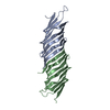
| ||||||||
|---|---|---|---|---|---|---|---|---|---|
| 1 |
| ||||||||
| Unit cell |
|
-Components
| #1: Protein | Mass: 20118.525 Da / Num. of mol.: 2 / Fragment: UNP residues 963-1138 Source method: isolated from a genetically manipulated source Source: (gene. exp.) #2: Water | ChemComp-HOH / |  Mass: 18.015 Da / Num. of mol.: 131 / Source method: isolated from a natural source / Formula: H2O Mass: 18.015 Da / Num. of mol.: 131 / Source method: isolated from a natural source / Formula: H2O |
|---|
-Experimental details
-Experiment
| Experiment | Method: X-RAY DIFFRACTION / Number of used crystals: 1 |
|---|
- Sample preparation
| Crystal | Density Matthews: 2.57 Å3/Da / Density % sol: 52.09 % Description: Squat boxes with one square side and bowed edges |
|---|---|
| Crystal grow | Temperature: 289 K / Method: vapor diffusion, sitting drop / pH: 7 / Details: 0.1 M HEPES, 15%(v/v) PEG 400, 0.2 M CaCl2 |
-Data collection
| Diffraction | Mean temperature: 100 K |
|---|---|
| Diffraction source | Source: SYNCHROTRON / Site: Diamond  / Beamline: I04 / Wavelength: 0.9763 Å / Beamline: I04 / Wavelength: 0.9763 Å |
| Detector | Type: DECTRIS PILATUS3 S 6M / Detector: PIXEL / Date: Dec 18, 2013 |
| Radiation | Monochromator: SILICON CRYSTAL / Protocol: SINGLE WAVELENGTH / Monochromatic (M) / Laue (L): M / Scattering type: x-ray |
| Radiation wavelength | Wavelength: 0.9763 Å / Relative weight: 1 |
| Reflection | Resolution: 1.85→49.57 Å / Num. obs: 35019 / % possible obs: 94.5 % / Redundancy: 9.2 % / CC1/2: 0.998 / Rmerge(I) obs: 0.083 / Rpim(I) all: 0.041 / Net I/σ(I): 11.3 |
| Reflection shell | Resolution: 1.85→1.89 Å / Redundancy: 6.8 % / Rmerge(I) obs: 2.887 / Mean I/σ(I) obs: 0.6 / Num. unique obs: 2208 / CC1/2: 0.586 / Rpim(I) all: 1.324 / % possible all: 99.2 |
- Processing
| Software |
| ||||||||||||||||||||||||||||||||||||||||||||||||||||||||||||||||||||||
|---|---|---|---|---|---|---|---|---|---|---|---|---|---|---|---|---|---|---|---|---|---|---|---|---|---|---|---|---|---|---|---|---|---|---|---|---|---|---|---|---|---|---|---|---|---|---|---|---|---|---|---|---|---|---|---|---|---|---|---|---|---|---|---|---|---|---|---|---|---|---|---|
| Refinement | Method to determine structure: SAD / Resolution: 1.859→49.571 Å / SU ML: 0.22 / Cross valid method: FREE R-VALUE / σ(F): 1.35 / Phase error: 28.86 / Stereochemistry target values: ML
| ||||||||||||||||||||||||||||||||||||||||||||||||||||||||||||||||||||||
| Solvent computation | Shrinkage radii: 0.9 Å / VDW probe radii: 1.11 Å / Solvent model: FLAT BULK SOLVENT MODEL | ||||||||||||||||||||||||||||||||||||||||||||||||||||||||||||||||||||||
| Refinement step | Cycle: LAST / Resolution: 1.859→49.571 Å
| ||||||||||||||||||||||||||||||||||||||||||||||||||||||||||||||||||||||
| Refine LS restraints |
| ||||||||||||||||||||||||||||||||||||||||||||||||||||||||||||||||||||||
| LS refinement shell |
| ||||||||||||||||||||||||||||||||||||||||||||||||||||||||||||||||||||||
| Refinement TLS params. | Method: refined / Origin x: 28.715 Å / Origin y: -24.2539 Å / Origin z: 249.4874 Å
| ||||||||||||||||||||||||||||||||||||||||||||||||||||||||||||||||||||||
| Refinement TLS group | Selection details: all |
