 Movie
Movie Controller

[English] 日本語
 Yorodumi
- PDB-6akg: Crystal structure of mouse claudin-3 P134G mutant in complex with... -
+ Open data

- Basic information
| Entry | Database: PDB / ID: 6akg | ||||||
|---|---|---|---|---|---|---|---|
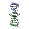
| Title | Crystal structure of mouse claudin-3 P134G mutant in complex with C-terminal fragment of Clostridium perfringens enterotoxin | ||||||
 Components Components |
| ||||||
 Keywords Keywords | MEMBRANE PROTEIN/TOXIN / Cell adhesion / Tight junction / MEMBRANE PROTEIN-TOXIN complex | ||||||
| Function / homology |  Function and homology information Function and homology informationcell junction maintenance / establishment of endothelial blood-brain barrier / calcium-independent cell-cell adhesion / response to Gram-positive bacterium / negative regulation of phosphate transmembrane transport / retinal pigment epithelium development / regulation of membrane permeability / cell-cell adhesion mediator activity / cell-cell junction maintenance / apicolateral plasma membrane ...cell junction maintenance / establishment of endothelial blood-brain barrier / calcium-independent cell-cell adhesion / response to Gram-positive bacterium / negative regulation of phosphate transmembrane transport / retinal pigment epithelium development / regulation of membrane permeability / cell-cell adhesion mediator activity / cell-cell junction maintenance / apicolateral plasma membrane / bicellular tight junction assembly / regulation of transepithelial transport / cell junction assembly / epithelial cell morphogenesis / regulation of cell morphogenesis / tight junction / apical junction complex / positive regulation of wound healing / lateral plasma membrane / bicellular tight junction / negative regulation of cell migration / cell-cell junction / cell junction / toxin activity / actin cytoskeleton organization / response to ethanol / response to hypoxia / cell adhesion / positive regulation of cell migration / negative regulation of cell population proliferation / negative regulation of gene expression / positive regulation of gene expression / structural molecule activity / protein-containing complex / extracellular region / membrane / identical protein binding / plasma membrane Similarity search - Function | ||||||
| Biological species |    Clostridium perfringens (bacteria) Clostridium perfringens (bacteria) | ||||||
| Method |  X-RAY DIFFRACTION / SYNCHROTRON / MOLECULAR REPLACEMENT / Resolution: 4.3 Å X-RAY DIFFRACTION / SYNCHROTRON / MOLECULAR REPLACEMENT / Resolution: 4.3 Å | ||||||
 Authors Authors | Nakamura, S. / Irie, K. / Fujiyoshi, Y. | ||||||
| Funding support |  Japan, 1items Japan, 1items
| ||||||
 Citation Citation | Journal: Nat Commun / Year: 2019 Title: Morphologic determinant of tight junctions revealed by claudin-3 structures. Authors: Nakamura, S. / Irie, K. / Tanaka, H. / Nishikawa, K. / Suzuki, H. / Saitoh, Y. / Tamura, A. / Tsukita, S. / Fujiyoshi, Y. | ||||||
| History |
|
- Structure visualization
| Structure viewer | Molecule: MolmilJmol/JSmol |
|---|
- Downloads & links
-Download
| PDBx/mmCIF format | 6akg.cif.gz | 126 KB | Display | PDBx/mmCIF format |
|---|---|---|---|---|
| PDB format | pdb6akg.ent.gz | 96.4 KB | Display | PDB format |
| PDBx/mmJSON format | 6akg.json.gz | Tree view | PDBx/mmJSON format | |
| Others |  Other downloads Other downloads |
-Validation report
| Arichive directory | https://data.pdbj.org/pub/pdb/validation_reports/ak/6akgftp://data.pdbj.org/pub/pdb/validation_reports/ak/6akg | HTTPS FTP |
|---|
-Related structure data
| Related structure data |  6akeC  6akfSC C: citing same article ( S: Starting model for refinement |
|---|---|
| Similar structure data |


-Links
 PDBj
PDBj
- Assembly
| Deposited unit | 
| ||||||||||||
|---|---|---|---|---|---|---|---|---|---|---|---|---|---|
| 1 | 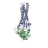
| ||||||||||||
| 2 | 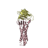
| ||||||||||||
| Unit cell |
|
-Components
| #1: Protein | Mass: 19866.439 Da / Num. of mol.: 2 / Fragment: UNP residues 1-183 / Mutation: P134A,C103A, C106A, C181A, C182A Source method: isolated from a genetically manipulated source Source: (gene. exp.)   Spodoptera frugiperda (fall armyworm) / References: UniProt: Q9Z0G9 Spodoptera frugiperda (fall armyworm) / References: UniProt: Q9Z0G9#2: Protein | Mass: 13329.830 Da / Num. of mol.: 2 / Fragment: UNP residues 203-319 / Mutation: S313A Source method: isolated from a genetically manipulated source Source: (gene. exp.) Clostridium perfringens (bacteria) / Gene: cpe / Production host: Has protein modification | Y | Sequence details | AUTHORS STATE THAT THE N-TERMINAL RESIDUES GHMASGS IN A AND C CHAINS ARE DERIVED FROM THE TEV ...AUTHORS STATE THAT THE N-TERMINAL RESIDUES GHMASGS IN A AND C CHAINS ARE DERIVED FROM THE TEV PROTEASE CLEAVAGE SITE AND LINKER AND THAT GLY201 AND SER202 IN B AND D CHAINS ARE DERIVED FROM THE THROMBIN CLEAVAGE SITE. | |
|---|
-Experimental details
-Experiment
| Experiment | Method: X-RAY DIFFRACTION / Number of used crystals: 1 |
|---|
- Sample preparation
| Crystal | Density Matthews: 5.13 Å3/Da / Density % sol: 76.02 % |
|---|---|
| Crystal grow | Temperature: 293.15 K / Method: vapor diffusion / pH: 8 / Details: HEPES, Magnesium nitrate, PEG 2000 MME |
-Data collection
| Diffraction | Mean temperature: 100 K |
|---|---|
| Diffraction source | Source: SYNCHROTRON / Site: SPring-8 / Beamline: BL41XU / Wavelength: 1 Å |
| Detector | Type: DECTRIS PILATUS3 6M / Detector: PIXEL / Date: Sep 24, 2016 |
| Radiation | Protocol: SINGLE WAVELENGTH / Monochromatic (M) / Laue (L): M / Scattering type: x-ray |
| Radiation wavelength | Wavelength: 1 Å / Relative weight: 1 |
| Reflection | Resolution: 4.3→49.34 Å / Num. obs: 9133 / % possible obs: 88.85 % / Redundancy: 6.7 % / Biso Wilson estimate: 214.57 Å2 / CC1/2: 0.999 / Rmerge(I) obs: 0.03868 / Rpim(I) all: 0.01638 / Rrim(I) all: 0.04212 / Net I/σ(I): 14.82 |
| Reflection shell | Resolution: 4.3→4.49 Å / Redundancy: 7 % / Rmerge(I) obs: 1.817 / Mean I/σ(I) obs: 1.31 / Num. unique obs: 490 / CC1/2: 0.689 / Rpim(I) all: 0.7385 / Rrim(I) all: 1.965 / % possible all: 53.84 |
- Processing
| Software |
| |||||||||||||||||||||||||||||||||||||||||||||||||||||||||||||||
|---|---|---|---|---|---|---|---|---|---|---|---|---|---|---|---|---|---|---|---|---|---|---|---|---|---|---|---|---|---|---|---|---|---|---|---|---|---|---|---|---|---|---|---|---|---|---|---|---|---|---|---|---|---|---|---|---|---|---|---|---|---|---|---|---|
| Refinement | Method to determine structure: MOLECULAR REPLACEMENT Starting model: 6AKF Resolution: 4.3→49.34 Å / SU ML: 0.8235 / Cross valid method: FREE R-VALUE / σ(F): 1.36 / Phase error: 52.4904
| |||||||||||||||||||||||||||||||||||||||||||||||||||||||||||||||
| Solvent computation | Shrinkage radii: 0.9 Å / VDW probe radii: 1.11 Å | |||||||||||||||||||||||||||||||||||||||||||||||||||||||||||||||
| Displacement parameters | Biso mean: 271.26 Å2 | |||||||||||||||||||||||||||||||||||||||||||||||||||||||||||||||
| Refinement step | Cycle: LAST / Resolution: 4.3→49.34 Å
| |||||||||||||||||||||||||||||||||||||||||||||||||||||||||||||||
| Refine LS restraints |
| |||||||||||||||||||||||||||||||||||||||||||||||||||||||||||||||
| LS refinement shell |
|
