ムービー
ムービー コントローラー


+ データを開く

- 基本情報
基本情報
| 登録情報 | データベース: EMDB / ID: EMD-1271 | |||||||||
|---|---|---|---|---|---|---|---|---|---|---|
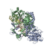
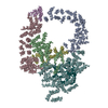
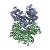
| タイトル | Structural model of full-length human Ku70-Ku80 heterodimer and its recognition of DNA and DNA-PKcs. | |||||||||
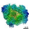
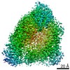
 マップデータ マップデータ | Frontal view of the Ku+DNA volume at a thresold of 2.5 | |||||||||
 試料 試料 |
| |||||||||
| 機能・相同性 | : / Ku70/Ku80 beta-barrel domain / nucleus 機能・相同性情報 機能・相同性情報 | |||||||||
| 生物種 |  Homo sapiens (ヒト) Homo sapiens (ヒト) | |||||||||
| 手法 | 単粒子再構成法 / ネガティブ染色法 / 解像度: 25.0 Å | |||||||||
 データ登録者 データ登録者 | Rivera-Calzada A / Spagnolo L / Pearl LH / Llorca O | |||||||||
 引用 引用 | ジャーナル: EMBO Rep / 年: 2007 タイトル: Structural model of full-length human Ku70-Ku80 heterodimer and its recognition of DNA and DNA-PKcs. 著者: Angel Rivera-Calzada / Laura Spagnolo / Laurence H Pearl / Oscar Llorca /  要旨: Recognition of DNA double-strand breaks during non-homologous end joining is carried out by the Ku70-Ku80 protein, a 150 kDa heterodimer that recruits the DNA repair kinase DNA-dependent protein ...Recognition of DNA double-strand breaks during non-homologous end joining is carried out by the Ku70-Ku80 protein, a 150 kDa heterodimer that recruits the DNA repair kinase DNA-dependent protein kinase catalytic subunit (DNA-PKcs) to the lesion. The atomic structure of a truncated Ku70-Ku80 was determined; however, the subunit-specific carboxy-terminal domain of Ku80--essential for binding to DNA-PKcs--was determined only in isolation, and the C-terminal domain of Ku70 was not resolved in its DNA-bound conformation. Both regions are conserved and mediate protein-protein interactions specific to mammals. Here, we reconstruct the three-dimensional structure of the human full-length Ku70-Ku80 dimer at 25 A resolution, alone and in complex with DNA, by using single-particle electron microscopy. We map the C-terminal regions of both subunits, and their conformational changes after DNA and DNA-PKcs binding to define a molecular model of the functions of these domains during DNA repair in the context of full-length Ku70-Ku80 protein. | |||||||||
| 履歴 |
|
- 構造の表示
構造の表示
| ムービー |
ムービービューア |
|---|---|
| 構造ビューア | EMマップ: SurfViewMolmilJmol/JSmol |
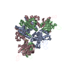
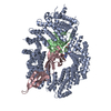
| 添付画像 |
 UCSF Chimera
- ダウンロードとリンク
-EMDBアーカイブ
| マップデータ | emd_1271.map.gz | 606.6 KB | EMDBマップデータ形式 | |
|---|---|---|---|---|
| ヘッダ (付随情報) | emd-1271-v30.xmlemd-1271.xml | 10.8 KB 10.8 KB | 表示 表示 | EMDBヘッダ |
| 画像 | 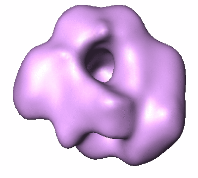 1271.gif 1271.gif | 44 KB | ||
| アーカイブディレクトリ |  http://ftp.pdbj.org/pub/emdb/structures/EMD-1271ftp://ftp.pdbj.org/pub/emdb/structures/EMD-1271 http://ftp.pdbj.org/pub/emdb/structures/EMD-1271ftp://ftp.pdbj.org/pub/emdb/structures/EMD-1271 | HTTPS FTP |
-検証レポート
| 文書・要旨 | emd_1271_validation.pdf.gz | 190.7 KB | 表示 | EMDB検証レポート |
|---|---|---|---|---|
| 文書・詳細版 | emd_1271_full_validation.pdf.gz | 189.8 KB | 表示 | |
| XML形式データ | emd_1271_validation.xml.gz | 5.4 KB | 表示 | |
| アーカイブディレクトリ | https://ftp.pdbj.org/pub/emdb/validation_reports/EMD-1271ftp://ftp.pdbj.org/pub/emdb/validation_reports/EMD-1271 | HTTPS FTP |
-関連構造データ


-リンク
| EMDBのページ | EMDB (EBI/PDBe) / EMDataResource |
|---|
-マップ
| ファイル | ダウンロード / ファイル: emd_1271.map.gz / 形式: CCP4 / 大きさ: 3.3 MB / タイプ: IMAGE STORED AS FLOATING POINT NUMBER (4 BYTES) | ||||||||||||||||||||||||||||||||||||||||||||||||||||||||||||||||||||
|---|---|---|---|---|---|---|---|---|---|---|---|---|---|---|---|---|---|---|---|---|---|---|---|---|---|---|---|---|---|---|---|---|---|---|---|---|---|---|---|---|---|---|---|---|---|---|---|---|---|---|---|---|---|---|---|---|---|---|---|---|---|---|---|---|---|---|---|---|---|
| 注釈 | Frontal view of the Ku+DNA volume at a thresold of 2.5 | ||||||||||||||||||||||||||||||||||||||||||||||||||||||||||||||||||||
| 投影像・断面図 | 画像のコントロール
画像は Spider により作成 | ||||||||||||||||||||||||||||||||||||||||||||||||||||||||||||||||||||
| ボクセルのサイズ | X=Y=Z: 2.12 Å | ||||||||||||||||||||||||||||||||||||||||||||||||||||||||||||||||||||
| 密度 |
| ||||||||||||||||||||||||||||||||||||||||||||||||||||||||||||||||||||
| 対称性 | 空間群: 1 | ||||||||||||||||||||||||||||||||||||||||||||||||||||||||||||||||||||
| 詳細 | EMDB XML:
CCP4マップ ヘッダ情報:
| ||||||||||||||||||||||||||||||||||||||||||||||||||||||||||||||||||||
 Z (Sec.)
 Y (Row.)
 X (Col.)


-添付データ
- 試料の構成要素
-全体 : Ku70-Ku80 heterodimer purified from HeLa cell nuclear extracts bo...
| 全体 | 名称: Ku70-Ku80 heterodimer purified from HeLa cell nuclear extracts bound to DNA |
|---|---|
| 要素 |
|
-超分子 #1000: Ku70-Ku80 heterodimer purified from HeLa cell nuclear extracts bo...
| 超分子 | 名称: Ku70-Ku80 heterodimer purified from HeLa cell nuclear extracts bound to DNA タイプ: sample / ID: 1000 集合状態: Heterodimer of Ku70 and Ku80 complexed bound to DNA Number unique components: 3 |
|---|---|
| 分子量 | 実験値: 152 KDa |
-分子 #1: Ku70
| 分子 | 名称: Ku70 / タイプ: protein_or_peptide / ID: 1 / コピー数: 1 / 組換発現: No |
|---|---|
| 由来(天然) | 生物種: Homo sapiens (ヒト) / 別称: Human / 細胞: HeLa / Organelle: Nucleus |
| 分子量 | 実験値: 70 KDa |
| 配列 | GO: GO: 0005624 / InterPro: Ku70/Ku80 beta-barrel domain |
-分子 #2: Ku80
| 分子 | 名称: Ku80 / タイプ: protein_or_peptide / ID: 2 / コピー数: 1 / 組換発現: No |
|---|---|
| 由来(天然) | 生物種: Homo sapiens (ヒト) / 別称: Human / 細胞: HeLa / Organelle: Nucleus |
| 分子量 | 実験値: 82 KDa |
| 配列 | GO: nucleus / InterPro: Ku70/Ku80 beta-barrel domain |
-分子 #3: DNA
| 分子 | 名称: DNA / タイプ: dna / ID: 3 / 詳細: 54 bp blunt-ended dsDNA 5' biotinilated / 分類: DNA / Structure: DOUBLE HELIX / Synthetic?: Yes |
|---|---|
| 配列 | 文字列: GGCCGCACGC GTCCACCATG GGGTACAACT ACGATCTAGC TTCATGCACC GGAC |
-実験情報
-構造解析
| 手法 | ネガティブ染色法 |
|---|---|
 解析 解析 | 単粒子再構成法 |
| 試料の集合状態 | particle |
-試料調製
| 濃度 | 0.068 mg/mL |
|---|---|
| 緩衝液 | 詳細: 50 mM Tris pH 7.5, 10% glycerol, 1mM DTT, 5 mM EDTA, 0.5mM Mg2Cl, 100mM NaCl, 20mM KCl |
| 染色 | タイプ: NEGATIVE 詳細: A few microliters of the DNA-bound Ku complexes were adsorbed to glow discharged carbon coated grids and negatively stained using 1% uranyl acetate. |
| グリッド | 詳細: 400 mesh Copper/Palladium grid |
| 凍結 | 凍結剤: NONE |
- 電子顕微鏡法
| 顕微鏡 | JEOL 1230 |
|---|---|
| アライメント法 | Legacy - 非点収差: correction with FFT and CCD camera |
| 詳細 | Microscope used: JEOL1230 |
| 日付 | 2005年4月20日 |
| 撮影 | カテゴリ: FILM / フィルム・検出器のモデル: KODAK 4489 FILM / デジタル化 - スキャナー: OTHER / デジタル化 - サンプリング間隔: 10 µm / 詳細: Scanner: MINOLTA Dimage Scan Multi Pro scanner / ビット/ピクセル: 16 |
| Tilt angle min | 0 |
| 電子線 | 加速電圧: 100 kV / 電子線源: TUNGSTEN HAIRPIN |
| 電子光学系 | 照射モード: FLOOD BEAM / 撮影モード: BRIGHT FIELD / Cs: 2.9 mm / 倍率(公称値): 50000 |
| 試料ステージ | 試料ホルダー: Eucentric / 試料ホルダーモデル: OTHER / Tilt angle max: 35 |
-画像解析
| 詳細 | Purified Ku and a ~10- fold molar excess of DNA were incubated at room temperature for 20 minutes. |
|---|---|
| 最終 再構成 | 想定した対称性 - 点群: C1 (非対称) / 解像度のタイプ: BY AUTHOR / 解像度: 25.0 Å / 解像度の算出法: FSC 0.5 CUT-OFF / ソフトウェア - 名称: EMAN / 使用した粒子像数: 8285 |
-原子モデル構築 1
| 初期モデル | PDB ID: |
|---|---|
| ソフトウェア | 名称: Situs |
| 詳細 | Protocol: Rigid Body. Fitting performed using Situs |
| 精密化 | プロトコル: RIGID BODY FIT / 当てはまり具合の基準: R-factor |