- EMDB-11999: M. pneumoniae 70S ribosome in complex with chloramphenicol obtain... -
+
データを開く
IDまたはキーワード:
読み込み中...
-
基本情報
登録情報
データベース: EMDB / ID: EMD-11999
タイトル
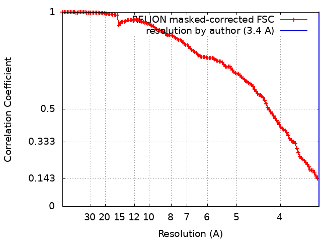
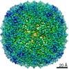
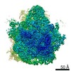
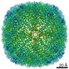
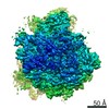
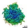
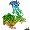
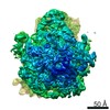
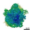
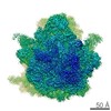
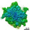
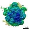
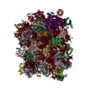
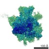
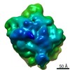
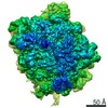
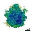
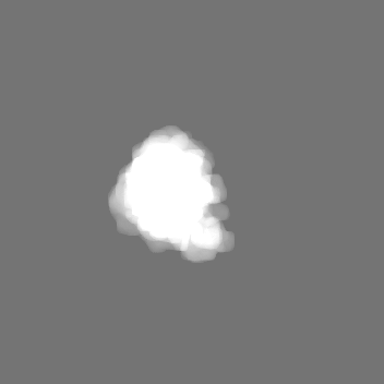
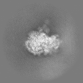
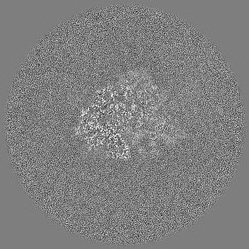
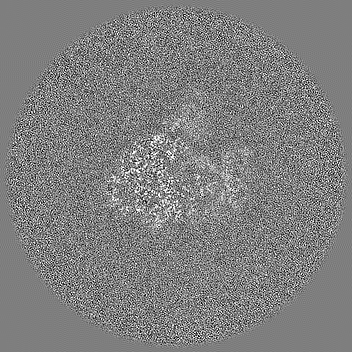
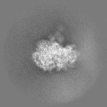
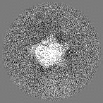
M. pneumoniae 70S ribosome in complex with chloramphenicol obtained from in situ data using M, focused refinement of 50S sub-unit
マップデータ
試料
複合体: 70S ribosome with chloramphenicol
機能・相同性
機能・相同性情報
large ribosomal subunit / transferase activity / 5S rRNA binding / ribosomal large subunit assembly / large ribosomal subunit rRNA binding / cytosolic large ribosomal subunit / cytoplasmic translation / tRNA binding / negative regulation of translation / rRNA binding ...large ribosomal subunit / transferase activity / 5S rRNA binding / ribosomal large subunit assembly / large ribosomal subunit rRNA binding / cytosolic large ribosomal subunit / cytoplasmic translation / tRNA binding / negative regulation of translation / rRNA binding / structural constituent of ribosome / ribosome / translation / ribonucleoprotein complex / response to antibiotic / mRNA binding / cytoplasm 類似検索 - 分子機能
Ribosomal protein L10, eubacterial, conserved site / Ribosomal protein L10 signature. / Ribosomal protein L10 / : / : / Ribosomal protein L11, bacterial-type / Ribosomal protein L31 type A / Ribosomal protein L31 signature. / Ribosomal protein L31 / Ribosomal protein L31 superfamily ...Ribosomal protein L10, eubacterial, conserved site / Ribosomal protein L10 signature. / Ribosomal protein L10 / : / : / Ribosomal protein L11, bacterial-type / Ribosomal protein L31 type A / Ribosomal protein L31 signature. / Ribosomal protein L31 / Ribosomal protein L31 superfamily / Ribosomal protein L31 / Ribosomal protein L11, conserved site / Ribosomal protein L11 signature. / Ribosomal protein L10-like domain superfamily / Ribosomal protein L10P / Ribosomal protein L10 / Ribosomal protein L16 signature 1. / Ribosomal protein L21, conserved site / Ribosomal protein L21 signature. / Ribosomal protein L16 signature 2. / Ribosomal protein L16, conserved site / Ribosomal protein L6, conserved site / Ribosomal protein L6 signature 1. / : / Ribosomal protein L9 signature. / Ribosomal protein L9, bacteria/chloroplast / Ribosomal protein L9, C-terminal / Ribosomal protein L9, C-terminal domain / Ribosomal protein L9, C-terminal domain superfamily / Ribosomal protein L11, N-terminal / Ribosomal protein L11, N-terminal domain / Ribosomal protein L11/L12 / Ribosomal protein L11, C-terminal / Ribosomal protein L11, C-terminal domain superfamily / Ribosomal protein L11/L12, N-terminal domain superfamily / Ribosomal protein L11/L12 / Ribosomal protein L11, RNA binding domain / Ribosomal protein L17 signature. / Ribosomal protein L36 signature. / Ribosomal protein L32p, bacterial type / Ribosomal protein L28/L24 superfamily / : / Ribosomal protein L9, N-terminal domain superfamily / Ribosomal protein L9 / Ribosomal protein L9, N-terminal / Ribosomal protein L9, N-terminal domain / Ribosomal protein L33, conserved site / Ribosomal protein L33 signature. / Ribosomal protein L35, conserved site / Ribosomal protein L35 signature. / Ribosomal protein L28 / Ribosomal protein L35, non-mitochondrial / Ribosomal protein L18, bacterial-type / : / Ribosomal protein L6, bacterial-type / Ribosomal protein L9/RNase H1, N-terminal / Ribosomal protein L5, bacterial-type / Ribosomal protein L36 / Ribosomal protein L36 superfamily / Ribosomal protein L36 / Ribosomal protein L19, conserved site / Ribosomal protein L19 signature. / Ribosomal protein L27, conserved site / Ribosomal protein L27 signature. / Ribosomal protein L20 signature. / Ribosomal protein L22, bacterial/chloroplast-type / Ribosomal protein L14P, bacterial-type / Ribosomal protein L34, conserved site / Ribosomal protein L34 signature. / Ribosomal protein L2, bacterial/organellar-type / Ribosomal protein L35 / Ribosomal protein L35 superfamily / Ribosomal protein L35 / Ribosomal protein L33 / Ribosomal protein L33 / Ribosomal protein L18 / Ribosomal L18 of archaea, bacteria, mitoch. and chloroplast / Ribosomal L28 family / Ribosomal protein L33 superfamily / Ribosomal protein L16 / Ribosomal protein L28/L24 / L28p-like / : / Ribosomal protein L27 / Ribosomal L27 protein / Ribosomal protein L20 / Ribosomal L32p protein family / Ribosomal protein L19 / Ribosomal protein L19 / Ribosomal protein L20 / Ribosomal protein L20, C-terminal / Ribosomal protein L19 superfamily / Ribosomal protein L21 / Ribosomal protein L32p / Ribosomal protein L17 / Ribosomal protein L17 superfamily / Ribosomal protein L17 / Ribosomal proteins 50S L24/mitochondrial 39S L24 / Ribosomal protein L21-like / L21-like superfamily 類似検索 - ドメイン・相同性
Large ribosomal subunit protein uL16 / Large ribosomal subunit protein bL36 / Large ribosomal subunit protein bL19 / Large ribosomal subunit protein bL28 / Large ribosomal subunit protein uL13 / Large ribosomal subunit protein bL32 / Large ribosomal subunit protein uL10 / Large ribosomal subunit protein bL35 / Large ribosomal subunit protein bL27 / Large ribosomal subunit protein bL9 ...Large ribosomal subunit protein uL16 / Large ribosomal subunit protein bL36 / Large ribosomal subunit protein bL19 / Large ribosomal subunit protein bL28 / Large ribosomal subunit protein uL13 / Large ribosomal subunit protein bL32 / Large ribosomal subunit protein uL10 / Large ribosomal subunit protein bL35 / Large ribosomal subunit protein bL27 / Large ribosomal subunit protein bL9 / Large ribosomal subunit protein uL11 / Large ribosomal subunit protein uL22 / Large ribosomal subunit protein uL2 / Large ribosomal subunit protein uL23 / Large ribosomal subunit protein uL4 / Large ribosomal subunit protein uL3 / Large ribosomal subunit protein bL34 / Large ribosomal subunit protein bL33A / Large ribosomal subunit protein bL31 / Large ribosomal subunit protein bL20 / Large ribosomal subunit protein bL21 / Large ribosomal subunit protein uL15 / Large ribosomal subunit protein uL18 / Large ribosomal subunit protein uL6 / Large ribosomal subunit protein uL5 / Large ribosomal subunit protein uL24 / Large ribosomal subunit protein uL14 / Large ribosomal subunit protein uL29 / Large ribosomal subunit protein bL17 類似検索 - 構成要素
ジャーナル: Nat Methods / 年: 2021 タイトル: Multi-particle cryo-EM refinement with M visualizes ribosome-antibiotic complex at 3.5 Å in cells. 著者: Dimitry Tegunov / Liang Xue / Christian Dienemann / Patrick Cramer / Julia Mahamid / 要旨: Cryo-electron microscopy (cryo-EM) enables macromolecular structure determination in vitro and inside cells. In addition to aligning individual particles, accurate registration of sample motion and ...Cryo-electron microscopy (cryo-EM) enables macromolecular structure determination in vitro and inside cells. In addition to aligning individual particles, accurate registration of sample motion and three-dimensional deformation during exposures are crucial for achieving high-resolution reconstructions. Here we describe M, a software tool that establishes a reference-based, multi-particle refinement framework for cryo-EM data and couples a comprehensive spatial deformation model to in silico correction of electron-optical aberrations. M provides a unified optimization framework for both frame-series and tomographic tilt-series data. We show that tilt-series data can provide the same resolution as frame-series data on a purified protein specimen, indicating that the alignment step no longer limits the resolution obtainable from tomographic data. In combination with Warp and RELION, M resolves to residue level a 70S ribosome bound to an antibiotic inside intact bacterial cells. Our work provides a computational tool that facilitates structural biology in cells.
EMPIAR-10499 (タイトル: Tilt series of native M. pneumoniae cells treated with chloramphenicol Data size: 83.8 Data #1: Unaligned tilt movies of M. pneumoniae [tilt series]) EMPIAR-10731 (タイトル: Locating Macromolecular Assemblies in Cells by 2D Template Matching with cisTEM Data size: 67.8 Data #1: 50S templates of Mycoplasma pneumoniae used for 2D and 3D template matching [reconstructed volumes] Data #2: Single-exposure micrographs of untilted Mycoplasma pneumoniae cells [micrographs - single frame] Data #3: Tomographic tilt series of Mycoplasma pneumoniae cells [tilt series] Data #4: Tomographic reconstructions of Mycoplasma pneumoniae cells [reconstructed volumes])
 ムービー
ムービー コントローラー


 データを開く
 基本情報
基本情報 マップデータ
 試料
試料 機能・相同性情報
 Mycoplasma pneumoniae (バクテリア)
 データ登録者
 ドイツ, 4件
  引用
引用 構造の表示
構造の表示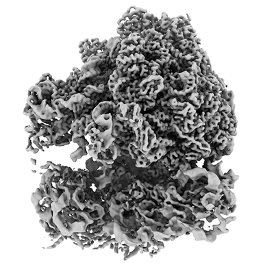
 ダウンロードとリンク
 emd_11999.png
 http://ftp.pdbj.org/pub/emdb/structures/EMD-11999


 Z (Sec.)
 Y (Row.)
 X (Col.)


 試料の構成要素
 解析
解析 電子顕微鏡法
 FIELD EMISSION GUN