+ Open data
- Basic information
| Entry | Database: EMDB / ID: EMD-11819 | |||||||||
|---|---|---|---|---|---|---|---|---|---|---|
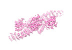
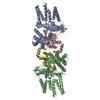
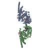
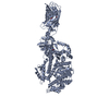
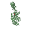
| Title | Structure of the body of the human TSC protein complex | |||||||||
 Map data Map data | Body of the human TSC protein complex | |||||||||
 Sample Sample |
| |||||||||
| Biological species |  Homo sapiens (human) Homo sapiens (human) | |||||||||
| Method | single particle reconstruction / cryo EM / Resolution: 4.2 Å | |||||||||
 Authors Authors | Ramlaul K / Fu W / Wu G / Aylett CHS | |||||||||
| Funding support |  United Kingdom, 1 items United Kingdom, 1 items
| |||||||||
 Citation Citation |  Journal: J Mol Biol / Year: 2021 Journal: J Mol Biol / Year: 2021Title: Architecture of the Tuberous Sclerosis Protein Complex. Authors: Kailash Ramlaul / Wencheng Fu / Hua Li / Natàlia de Martin Garrido / Lin He / Manjari Trivedi / Wei Cui / Christopher H S Aylett / Geng Wu /   Abstract: The Tuberous Sclerosis Complex (TSC) protein complex (TSCC), comprising TSC1, TSC2, and TBC1D7, is widely recognised as a key integration hub for cell growth and intracellular stress signals upstream ...The Tuberous Sclerosis Complex (TSC) protein complex (TSCC), comprising TSC1, TSC2, and TBC1D7, is widely recognised as a key integration hub for cell growth and intracellular stress signals upstream of the mammalian target of rapamycin complex 1 (mTORC1). The TSCC negatively regulates mTORC1 by acting as a GTPase-activating protein (GAP) towards the small GTPase Rheb. Both human TSC1 and TSC2 are important tumour suppressors, and mutations in them underlie the disease tuberous sclerosis. We used single-particle cryo-EM to reveal the organisation and architecture of the complete human TSCC. We show that TSCC forms an elongated scorpion-like structure, consisting of a central "body", with a "pincer" and a "tail" at the respective ends. The "body" is composed of a flexible TSC2 HEAT repeat dimer, along the surface of which runs the TSC1 coiled-coil backbone, breaking the symmetry of the dimer. Each end of the body is structurally distinct, representing the N- and C-termini of TSC1; a "pincer" is formed by the highly flexible N-terminal TSC1 core domains and a barbed "tail" makes up the TSC1 coiled-coil-TBC1D7 junction. The TSC2 GAP domain is found abutting the centre of the body on each side of the dimerisation interface, poised to bind a pair of Rheb molecules at a similar separation to the pair in activated mTORC1. Our architectural dissection reveals the mode of association and topology of the complex, casts light on the recruitment of Rheb to the TSCC, and also hints at functional higher order oligomerisation, which has previously been predicted to be important for Rheb-signalling suppression. | |||||||||
| History |
|
- Structure visualization
| Movie |
 Movie viewer Movie viewer |
|---|---|
| Structure viewer | EM map:  SurfView SurfView Molmil Molmil Jmol/JSmol Jmol/JSmol |
| Supplemental images |
- Downloads & links
-EMDB archive
| Map data |  emd_11819.map.gz emd_11819.map.gz | 10.7 MB |  EMDB map data format EMDB map data format | |
|---|---|---|---|---|
| Header (meta data) |  emd-11819-v30.xml emd-11819-v30.xml emd-11819.xml emd-11819.xml | 22.6 KB 22.6 KB | Display Display |  EMDB header EMDB header |
| Images |  emd_11819.png emd_11819.png | 111.7 KB | ||
| Others |  emd_11819_half_map_1.map.gz emd_11819_half_map_1.map.gz emd_11819_half_map_2.map.gz emd_11819_half_map_2.map.gz | 8 MB 8 MB | ||
| Archive directory |  http://ftp.pdbj.org/pub/emdb/structures/EMD-11819 http://ftp.pdbj.org/pub/emdb/structures/EMD-11819 ftp://ftp.pdbj.org/pub/emdb/structures/EMD-11819 ftp://ftp.pdbj.org/pub/emdb/structures/EMD-11819 | HTTPS FTP |
-Validation report
| Summary document |  emd_11819_validation.pdf.gz emd_11819_validation.pdf.gz | 365 KB | Display |  EMDB validaton report EMDB validaton report |
|---|---|---|---|---|
| Full document |  emd_11819_full_validation.pdf.gz emd_11819_full_validation.pdf.gz | 364.1 KB | Display | |
| Data in XML |  emd_11819_validation.xml.gz emd_11819_validation.xml.gz | 8.3 KB | Display | |
| Arichive directory |  https://ftp.pdbj.org/pub/emdb/validation_reports/EMD-11819 https://ftp.pdbj.org/pub/emdb/validation_reports/EMD-11819 ftp://ftp.pdbj.org/pub/emdb/validation_reports/EMD-11819 ftp://ftp.pdbj.org/pub/emdb/validation_reports/EMD-11819 | HTTPS FTP |
-Related structure data
| Related structure data | C: citing same article ( |
|---|---|
| Similar structure data | |
| EM raw data |  EMPIAR-10704 (Title: TSC complex particles / Data size: 93.7 EMPIAR-10704 (Title: TSC complex particles / Data size: 93.7 Data #1: Dose weighted micrographs [micrographs - single frame])  EMPIAR-10715 (Title: TSC complex particles / Data size: 13.1 TB / Data #1: TSC particle frames [micrographs - multiframe]) EMPIAR-10715 (Title: TSC complex particles / Data size: 13.1 TB / Data #1: TSC particle frames [micrographs - multiframe]) |
- Links
Links
| EMDB pages |  EMDB (EBI/PDBe) / EMDB (EBI/PDBe) /  EMDataResource EMDataResource |
|---|
- Map
Map
| File |  Download / File: emd_11819.map.gz / Format: CCP4 / Size: 11.4 MB / Type: IMAGE STORED AS FLOATING POINT NUMBER (4 BYTES) Download / File: emd_11819.map.gz / Format: CCP4 / Size: 11.4 MB / Type: IMAGE STORED AS FLOATING POINT NUMBER (4 BYTES) | ||||||||||||||||||||||||||||||||||||||||||||||||||||||||||||||||||||
|---|---|---|---|---|---|---|---|---|---|---|---|---|---|---|---|---|---|---|---|---|---|---|---|---|---|---|---|---|---|---|---|---|---|---|---|---|---|---|---|---|---|---|---|---|---|---|---|---|---|---|---|---|---|---|---|---|---|---|---|---|---|---|---|---|---|---|---|---|---|
| Annotation | Body of the human TSC protein complex | ||||||||||||||||||||||||||||||||||||||||||||||||||||||||||||||||||||
| Projections & slices | Image control
Images are generated by Spider. | ||||||||||||||||||||||||||||||||||||||||||||||||||||||||||||||||||||
| Voxel size | X=Y=Z: 1.35 Å | ||||||||||||||||||||||||||||||||||||||||||||||||||||||||||||||||||||
| Density |
| ||||||||||||||||||||||||||||||||||||||||||||||||||||||||||||||||||||
| Symmetry | Space group: 1 | ||||||||||||||||||||||||||||||||||||||||||||||||||||||||||||||||||||
| Details | EMDB XML:
CCP4 map header:
| ||||||||||||||||||||||||||||||||||||||||||||||||||||||||||||||||||||
-Supplemental data
-Half map: Body of the human TSC protein complex
| File | emd_11819_half_map_1.map | ||||||||||||
|---|---|---|---|---|---|---|---|---|---|---|---|---|---|
| Annotation | Body of the human TSC protein complex | ||||||||||||
| Projections & Slices |
| ||||||||||||
| Density Histograms |
-Half map: Body of the human TSC protein complex
| File | emd_11819_half_map_2.map | ||||||||||||
|---|---|---|---|---|---|---|---|---|---|---|---|---|---|
| Annotation | Body of the human TSC protein complex | ||||||||||||
| Projections & Slices |
| ||||||||||||
| Density Histograms |
- Sample components
-Entire : Body of the human TSC protein complex
| Entire | Name: Body of the human TSC protein complex |
|---|---|
| Components |
|
-Supramolecule #1: Body of the human TSC protein complex
| Supramolecule | Name: Body of the human TSC protein complex / type: complex / ID: 1 / Parent: 0 / Macromolecule list: all |
|---|---|
| Source (natural) | Organism:  Homo sapiens (human) Homo sapiens (human) |
| Recombinant expression | Organism:  Homo sapiens (human) Homo sapiens (human) |
| Molecular weight | Experimental: 700 KDa |
-Macromolecule #1: Hamartin
| Macromolecule | Name: Hamartin / type: protein_or_peptide / ID: 1 / Enantiomer: LEVO |
|---|---|
| Source (natural) | Organism:  Homo sapiens (human) Homo sapiens (human) |
| Recombinant expression | Organism:  Homo sapiens (human) Homo sapiens (human) |
| Sequence | String: MAQQANVGEL LAMLDSPMLG VRDDVTAVFK ENLNSDRGPM LVNTLVDYYL ETSSQPALHI LTTLQEPHD KHLLDRINEY VGKAATRLSI LSLLGHVIRL QPSWKHKLSQ APLLPSLLKC L KMDTDVVV LTTGVLVLIT MLPMIPQSGK QHLLDFFDIF GRLSSWCLKK ...String: MAQQANVGEL LAMLDSPMLG VRDDVTAVFK ENLNSDRGPM LVNTLVDYYL ETSSQPALHI LTTLQEPHD KHLLDRINEY VGKAATRLSI LSLLGHVIRL QPSWKHKLSQ APLLPSLLKC L KMDTDVVV LTTGVLVLIT MLPMIPQSGK QHLLDFFDIF GRLSSWCLKK PGHVAEVYLV HL HASVYAL FHRLYGMYPC NFVSFLRSHY SMKENLETFE EVVKPMMEHV RIHPELVTGS KDH ELDPRR WKRLETHDVV IECAKISLDP TEASYEDGYS VSHQISARFP HRSADVTTSP YADT QNSYG CATSTPYSTS RLMLLNMPGQ LPQTLSSPST RLITEPPQAT LWSPSMVCGM TTPPT SPGN VPPDLSHPYS KVFGTTAGGK GTPLGTPATS PPPAPLCHSD DYVHISLPQA TVTPPR KEE RMDSARPCLH RQHHLLNDRG SEEPPGSKGS VTLSDLPGFL GDLASEEDSI EKDKEEA AI SRELSEITTA EAEPVVPRGG FDSPFYRDSL PGSQRKTHSA ASSSQGASVN PEPLHSSL D KLGPDTPKQA FTPIDLPCGS ADESPAGDRE CQTSLETSIF TPSPCKIPPP TRVGFGSGQ PPPYDHLFEV ALPKTAHHFV IRKTEELLKK AKGNTEEDGV PSTSPMEVLD RLIQQGADAH SKELNKLPL PSKSVDWTHF GGSPPSDEIR TLRDQLLLLH NQLLYERFKR QQHALRNRRL L RKVIKAAA LEEHNAAMKD QLKLQEKDIQ MWKVSLQKEQ ARYNQLQEQR DTMVTKLHSQ IR QLQHDRE EFYNQSQELQ TKLEDCRNMI AELRIELKKA NNKVCHTELL LSQVSQKLSN SES VQQQME FLNRQLLVLG EVNELYLEQL QNKHSDTTKE VEMMKAAYRK ELEKNRSHVL QQTQ RLDTS QKRILELESH LAKKDHLLLE QKKYLEDVKL QARGQLQAAE SRYEAQKRIT QVFEL EILD LYGRLEKDGL LKKLEEEKAE AAEAAEERLD CCNDGCSDSM VGHNEEASGH NGETKT PRP SSARGSSGSR GGGGSSSSSS ELSTPEKPPH QRAGPFSSRW ETTMGEASAS IPTTVGS LP SSKSFLGMKA RELFRNKSES QCDEDGMTSS LSESLKTELG KDLGVEAKIP LNLDGPHP S PPTPDSVGQL HIMDYNETHH EHS |
-Macromolecule #2: Tuberin
| Macromolecule | Name: Tuberin / type: protein_or_peptide / ID: 2 / Enantiomer: LEVO |
|---|---|
| Source (natural) | Organism:  Homo sapiens (human) Homo sapiens (human) |
| Recombinant expression | Organism:  Homo sapiens (human) Homo sapiens (human) |
| Sequence | String: MAKPTSKDSG LKEKFKILLG LGTPRPNPRS AEGKQTEFII TAEILRELSM ECGLNNRIRM IGQICEVAK TKKFEEHAVE ALWKAVADLL QPERPLEARH AVLALLKAIV QGQGERLGVL R ALFFKVIK DYPSNEDLHE RLEVFKALTD NGRHITYLEE ELADFVLQWM ...String: MAKPTSKDSG LKEKFKILLG LGTPRPNPRS AEGKQTEFII TAEILRELSM ECGLNNRIRM IGQICEVAK TKKFEEHAVE ALWKAVADLL QPERPLEARH AVLALLKAIV QGQGERLGVL R ALFFKVIK DYPSNEDLHE RLEVFKALTD NGRHITYLEE ELADFVLQWM DVGLSSEFLL VL VNLVKFN SCYLDEYIAR MVQMICLLCV RTASSVDIEV SLQVLDAVVC YNCLPAESLP LFI VTLCRT INVKELCEPC WKLMRNLLGT HLGHSAIYNM CHLMEDRAYM EDAPLLRGAV FFVG MALWG AHRLYSLRNS PTSVLPSFYQ AMACPNEVVS YEIVLSITRL IKKYRKELQV VAWDI LLNI IERLLQQLQT LDSPELRTIV HDLLTTVEEL CDQNEFHGSQ ERYFELVERC ADQRPE SSL LNLISYRAQS IHPAKDGWIQ NLQALMERFF RSESRGAVRI KVLDVLSFVL LINRQFY EE ELINSVVISQ LSHIPEDKDH QVRKLATQLL VDLAEGCHTH HFNSLLDIIE KVMARSLS P PPELEERDVA AYSASLEDVK TAVLGLLVIL QTKLYTLPAS HATRVYEMLV SHIQLHYKH SYTLPIASSI RLQAFDFLLL LRADSLHRLG LPNKDGVVRF SPYCVCDYME PERGSEKKTS GPLSPPTGP PGPAPAGPAV RLGSVPYSLL FRVLLQCLKQ ESDWKVLKLV LGRLPESLRY K VLIFTSPC SVDQLCSALC SMLSGPKTLE RLRGAPEGFS RTDLHLAVVP VLTALISYHN YL DKTKQRE MVYCLEQGLI HRCASQCVVA LSICSVEMPD IIIKALPVLV VKLTHISATA SMA VPLLEF LSTLARLPHL YRNFAAEQYA SVFAISLPYT NPSKFNQYIV CLAHHVIAMW FIRC RLPFR KDFVPFITKG LRSNVLLSFD DTPEKDSFRA RSTSLNERPK SLRIARPPKQ GLNNS PPVK EFKESSAAEA FRCRSISVSE HVVRSRIQTS LTSASLGSAD ENSVAQADDS LKNLHL ELT ETCLDMMARY VFSNFTAVPK RSPVGEFLLA GGRTKTWLVG NKLVTVTTSV GTGTRSL LG LDSGELQSGP ESSSSPGVHV RQTKEAPAKL ESQAGQQVSR GARDRVRSMS GGHGLRVG A LDVPASQFLG SATSPGPRTA PAAKPEKASA GTRVPVQEKT NLAAYVPLLT QGWAEILVR RPTGNTSWLM SLENPLSPFS SDINNMPLQE LSNALMAAER FKEHRDTALY KSLSVPAAST AKPPPLPRS NTVASFSSLY QSSCQGQLHR SVSWADSAVV MEEGSPGEVP VLVEPPGLED V EAALGMDR RTDAYSRSSS VSSQEEKSLH AEELVGRGIP IERVVSSEGG RPSVDLSFQP SQ PLSKSSS SPELQTLQDI LGDPGDKADV GRLSPEVKAR SQSGTLDGES AAWSASGEDS RGQ PEGPLP SSSPRSPSGL RPRGYTISDS APSRRGKRVE RDALKSRATA SNAEKVPGIN PSFV FLQLY HSPFFGDESN KPILLPNESQ SFERSVQLLD QIPSYDTHKI AVLYVGEGQS NSELA ILSN EHGSYRYTEF LTGLGRLIEL KDCQPDKVYL GGLDVCGEDG QFTYCWHDDI MQAVFH IAT LMPTKDVDKH RCDKKRHLGN DFVSIVYNDS GEDFKLGTIK GQFNFVHVIV TPLDYEC NL VSLQCRKDME GLVDTSVAKI VSDRNLPFVA RQMALHANMA SQVHHSRSNP TDIYPSKW I ARLRHIKRLR QRICEEAAYS NPSLPLVHPP SHSKAPAQTP AEPTPGYEVG QRKRLISSV EDFTEFV |
-Macromolecule #3: TBC1D7
| Macromolecule | Name: TBC1D7 / type: protein_or_peptide / ID: 3 / Enantiomer: LEVO |
|---|---|
| Source (natural) | Organism:  Homo sapiens (human) Homo sapiens (human) |
| Recombinant expression | Organism:  Homo sapiens (human) Homo sapiens (human) |
| Sequence | String: MTEDSQRNFR SVYYEKVGFR GVEEKKSLEI LLKDDRLDTE KLCTFSQRFP LPSMYRALVW KVLLGILPP HHESHAKVMM YRKEQYLDVL HALKVVRFVS DATPQAEVYL RMYQLESGKL P RSPSFPLE PDDEVFLAIA KAMEEMVEDS VDCYWITRRF VNQLNTKYRD ...String: MTEDSQRNFR SVYYEKVGFR GVEEKKSLEI LLKDDRLDTE KLCTFSQRFP LPSMYRALVW KVLLGILPP HHESHAKVMM YRKEQYLDVL HALKVVRFVS DATPQAEVYL RMYQLESGKL P RSPSFPLE PDDEVFLAIA KAMEEMVEDS VDCYWITRRF VNQLNTKYRD SLPQLPKAFE QY LNLEDGR LLTHLRMCSA APKLPYDLWF KRCFAGCLPE SSLQRVWDKV VSGSCKILVF VAV EILLTF KIKVMALNSA EKITKFLENI PQDSSDAIVS KAIDLWHKHC GTPVHSS |
-Experimental details
-Structure determination
| Method | cryo EM |
|---|---|
 Processing Processing | single particle reconstruction |
| Aggregation state | particle |
- Sample preparation
| Concentration | 0.2 mg/mL |
|---|---|
| Buffer | pH: 7.6 Details: 25 mM K-HEPES, pH 7.6, 175 mM KCl or 150 mM LiCl, 1 mM TCEP, and 0.5 uM EDTA |
| Grid | Model: Quantifoil R2/1 / Material: COPPER / Mesh: 300 / Support film - #0 - Film type ID: 1 / Support film - #0 - Material: CARBON / Support film - #0 - topology: HOLEY ARRAY / Support film - #1 - Film type ID: 2 / Support film - #1 - Material: GRAPHENE OXIDE / Support film - #1 - topology: CONTINUOUS / Pretreatment - Type: PLASMA CLEANING / Pretreatment - Atmosphere: AIR |
| Vitrification | Cryogen name: ETHANE / Chamber humidity: 96 % / Chamber temperature: 278 K / Instrument: FEI VITROBOT MARK IV / Details: 2 seconds blotting. |
- Electron microscopy
| Microscope | FEI TITAN KRIOS |
|---|---|
| Specialist optics | Energy filter - Name: GIF Bioquantum / Energy filter - Slit width: 70 eV |
| Image recording | Film or detector model: GATAN K2 SUMMIT (4k x 4k) / Detector mode: SUPER-RESOLUTION / Digitization - Dimensions - Width: 3838 pixel / Digitization - Dimensions - Height: 3710 pixel / Digitization - Sampling interval: 5.0 µm / Number real images: 5387 / Average electron dose: 40.0 e/Å2 |
| Electron beam | Acceleration voltage: 300 kV / Electron source:  FIELD EMISSION GUN FIELD EMISSION GUN |
| Electron optics | C2 aperture diameter: 70.0 µm / Illumination mode: FLOOD BEAM / Imaging mode: BRIGHT FIELD / Cs: 2.7 mm / Nominal defocus max: 3.0 µm / Nominal defocus min: 1.0 µm |
| Sample stage | Specimen holder model: FEI TITAN KRIOS AUTOGRID HOLDER / Cooling holder cryogen: NITROGEN |
| Experimental equipment |  Model: Titan Krios / Image courtesy: FEI Company |
+ Image processing
-Atomic model buiding 1
| Refinement | Protocol: RIGID BODY FIT |
|---|
 Movie
Movie Controller







 Z (Sec.)
 Y (Row.)
 X (Col.)


