[English] 日本語
Yorodumi
- EMDB-11700: HBV pgRNA T=4 NCP icosahedral symmetry -

+
Open data


ID or keywords:

Loading...

-
Basic information

Entry
Database: EMDB / ID: EMD-11700
TitleHBV pgRNA T=4 NCP icosahedral symmetry
Map dataHBV T=4 NCP icosahedral symmetry
Sample
  • Virus: Hepatitis B virus
    • Protein or peptide: Capsid protein
KeywordsHBV / VLP / pgRNA / VIRUS LIKE PARTICLE
Function / homology
Function and homology information


virus-mediated perturbation of host defense response => GO:0019049 / microtubule-dependent intracellular transport of viral material towards nucleus / T=4 icosahedral viral capsid / : / viral penetration into host nucleus / host cell cytoplasm / symbiont entry into host cell / structural molecule activity / DNA binding / RNA binding / extracellular region
Similarity search - Function
Hepatitis B virus, capsid N-terminal / Hepatitis core protein, putative zinc finger / Hepatitis core antigen / Viral capsid core domain supefamily, Hepatitis B virus / Hepatitis core antigen
Similarity search - Domain/homology
Biological speciesHepatitis B virus
Methodsingle particle reconstruction / cryo EM / Resolution: 3.2 Å
AuthorsPatel N / Clark S
Funding support United Kingdom, 2 items
OrganizationGrant numberCountry
Medical Research Council (MRC, United Kingdom)MRF-044-0002-RG-PATEL; MR/N021517/1 United Kingdom
Wellcome TrustJoint Investigator Award Nos. 110145 & 110146; 089311/Z/09/Z; 090932/Z/09/Z & 106692 United Kingdom
Citation
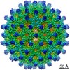
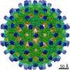
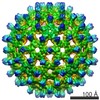
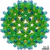
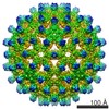
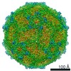
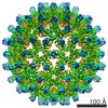
Journal: Commun Biol / Year: 2021
Title: In vitro functional analysis of gRNA sites regulating assembly of hepatitis B virus.
Authors: Nikesh Patel / Sam Clark / Eva U Weiß / Carlos P Mata / Jen Bohon / Erik R Farquhar / Daniel P Maskell / Neil A Ranson / Reidun Twarock / Peter G Stockley /
Abstract: The roles of RNA sequence/structure motifs, Packaging Signals (PSs), for regulating assembly of an HBV genome transcript have been investigated in an efficient in vitro assay containing only core ...The roles of RNA sequence/structure motifs, Packaging Signals (PSs), for regulating assembly of an HBV genome transcript have been investigated in an efficient in vitro assay containing only core protein (Cp) and RNA. Variants of three conserved PSs, within the genome of a strain not used previously, preventing correct presentation of a Cp-recognition loop motif are differentially deleterious for assembly of nucleocapsid-like particles (NCPs). Cryo-electron microscopy reconstruction of the T = 4 NCPs formed with the wild-type gRNA transcript, reveal that the interior of the Cp shell is in contact with lower resolution density, potentially encompassing the arginine-rich protein domains and gRNA. Symmetry relaxation followed by asymmetric reconstruction reveal that such contacts are made at every symmetry axis. We infer from their regulation of assembly that some of these contacts would involve gRNA PSs, and confirmed this by X-ray RNA footprinting. Mutation of the ε stem-loop in the gRNA, where polymerase binds in vivo, produces a poor RNA assembly substrate with Cp alone, largely due to alterations in its conformation. The results show that RNA PSs regulate assembly of HBV genomic transcripts in vitro, and therefore may play similar roles in vivo, in concert with other molecular factors.
History
DepositionSep 7, 2020-
Header (metadata) releaseOct 27, 2021-
Map releaseOct 27, 2021-
UpdateJul 10, 2024-
Current statusJul 10, 2024Processing site: PDBe / Status: Released

-
Structure visualization

Movie
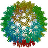
  • Surface view with section colored by density value
  • Surface level: 0.026
  • Imaged by UCSF Chimera
  • Download
  • Surface view colored by radius
  • Surface level: 0.026
  • Imaged by UCSF Chimera
  • Download
  • Surface view with fitted model
  • Atomic models: PDB-7abl
  • Surface level: 0.026
  • Imaged by UCSF Chimera
  • Download
  • Simplified surface model + fitted atomic model
  • Atomic modelsPDB-7abl
  • Imaged by Jmol
  • Download
Movie viewer
Structure viewerEM map:
SurfViewMolmilJmol/JSmol
Supplemental images

Downloads & links

-
Map

FileDownload / File: emd_11700.map.gz / Format: CCP4 / Size: 209.3 MB / Type: IMAGE STORED AS FLOATING POINT NUMBER (4 BYTES)
AnnotationHBV T=4 NCP icosahedral symmetry
Voxel sizeX=Y=Z: 1.065 Å
Density
Contour LevelBy AUTHOR: 0.026 / Movie #1: 0.026
Minimum - Maximum-0.13050236 - 0.20400919
Average (Standard dev.)0.0009919029 (±0.013941099)
SymmetrySpace group: 1
Details

EMDB XML:

Map geometry
Axis orderXYZ
Origin-190-190-190
Dimensions380380380
Spacing380380380
CellA=B=C: 404.7 Å
α=β=γ: 90.0 °

CCP4 map header:

modeImage stored as Reals
Å/pix. X/Y/Z1.0651.0651.065
M x/y/z380380380
origin x/y/z0.0000.0000.000
length x/y/z404.700404.700404.700
α/β/γ90.00090.00090.000
MAP C/R/S123
start NC/NR/NS-190-190-190
NC/NR/NS380380380
D min/max/mean-0.1310.2040.001

-
Supplemental data

-
Sample components

-
Entire : Hepatitis B virus

EntireName: Hepatitis B virus
Components
  • Virus: Hepatitis B virus
    • Protein or peptide: Capsid protein

-
Supramolecule #1: Hepatitis B virus

SupramoleculeName: Hepatitis B virus / type: virus / ID: 1 / Parent: 0 / Macromolecule list: all / NCBI-ID: 10407 / Sci species name: Hepatitis B virus / Virus type: VIRUS-LIKE PARTICLE / Virus isolate: OTHER / Virus enveloped: No / Virus empty: No
Virus shellShell ID: 1 / Diameter: 342.0 Å / T number (triangulation number): 4

-
Macromolecule #1: Capsid protein

MacromoleculeName: Capsid protein / type: protein_or_peptide / ID: 1 / Number of copies: 4 / Enantiomer: LEVO
Source (natural)Organism: Hepatitis B virus
Molecular weightTheoretical: 21.125199 KDa
Recombinant expressionOrganism: Escherichia coli (E. coli)
SequenceString:
MDIDPYKEFG ASVELLSFLP SDFFPSIRDL LDTASALYRE ALESPEHCSP HHTALRQAIL CWGELMNLAT WVGSNLEDPA SRELVVSYV NVNMGLKIRQ LLWFHISCLT FGRETVLEYL VSFGVWIRTP PAYRPPNAPI LSTLPETTVV RRRGRSPRRR T PSPRRRRS QSPRRRRSQS RESQC

UniProtKB: Capsid protein

-
Experimental details

-
Structure determination

Methodcryo EM
Processingsingle particle reconstruction
Aggregation stateparticle

-
Sample preparation

BufferpH: 7.5
GridModel: Quantifoil / Support film - Material: CARBON / Support film - topology: LACEY / Pretreatment - Type: GLOW DISCHARGE / Pretreatment - Time: 30 sec.
VitrificationCryogen name: ETHANE / Chamber humidity: 95 % / Chamber temperature: 277 K / Instrument: LEICA EM GP

-
Electron microscopy

MicroscopeFEI TITAN KRIOS
Image recordingFilm or detector model: FEI FALCON III (4k x 4k) / Detector mode: INTEGRATING / Number grids imaged: 1 / Number real images: 2658 / Average exposure time: 1.5 sec. / Average electron dose: 53.1 e/Å2
Electron beamAcceleration voltage: 300 kV / Electron source: FIELD EMISSION GUN
Electron opticsC2 aperture diameter: 70.0 µm / Illumination mode: FLOOD BEAM / Imaging mode: OTHER / Cs: 2.7 mm / Nominal defocus max: 2.5 µm / Nominal defocus min: 0.75 µm / Nominal magnification: 75000
Sample stageSpecimen holder model: FEI TITAN KRIOS AUTOGRID HOLDER / Cooling holder cryogen: NITROGEN
Experimental equipment
Model: Titan Krios / Image courtesy: FEI Company

+
Image processing

Particle selectionNumber selected: 150667
Startup modelType of model: EMDB MAP
Final reconstructionApplied symmetry - Point group: I (icosahedral) / Algorithm: FOURIER SPACE / Resolution.type: BY AUTHOR / Resolution: 3.2 Å / Resolution method: FSC 0.143 CUT-OFF / Software - Name: RELION (ver. 3.0) / Number images used: 104337
Initial angle assignmentType: MAXIMUM LIKELIHOOD / Software - Name: RELION (ver. 3.0)
Final angle assignmentType: MAXIMUM LIKELIHOOD / Software - Name: RELION (ver. 3.0)

-
Atomic model buiding 1

Initial modelPDB ID:

Chain - Source name: PDB / Chain - Initial model type: experimental model
RefinementSpace: REAL / Protocol: OTHER / Overall B value: 158
Output model

PDB-7abl:
HBV pgRNA T=4 NCP icosahedral symmetry

+
About Yorodumi

-
News

-
Feb 9, 2022. New format data for meta-information of EMDB entries

New format data for meta-information of EMDB entries

  • Version 3 of the EMDB header file is now the official format.
  • The previous official version 1.9 will be removed from the archive.

Related info.:EMDB header

External links:wwPDB to switch to version 3 of the EMDB data model

-
Aug 12, 2020. Covid-19 info

Covid-19 info

URL: https://pdbjlvh1.pdbj.org/emnavi/covid19.php

New page: Covid-19 featured information page in EM Navigator.

Related info.:Covid-19 info / Mar 5, 2020. Novel coronavirus structure data

+
Mar 5, 2020. Novel coronavirus structure data

Novel coronavirus structure data

Related info.:Yorodumi Speices / Aug 12, 2020. Covid-19 info

External links:COVID-19 featured content - PDBj / Molecule of the Month (242):Coronavirus Proteases

+
Jan 31, 2019. EMDB accession codes are about to change! (news from PDBe EMDB page)

EMDB accession codes are about to change! (news from PDBe EMDB page)

  • The allocation of 4 digits for EMDB accession codes will soon come to an end. Whilst these codes will remain in use, new EMDB accession codes will include an additional digit and will expand incrementally as the available range of codes is exhausted. The current 4-digit format prefixed with “EMD-” (i.e. EMD-XXXX) will advance to a 5-digit format (i.e. EMD-XXXXX), and so on. It is currently estimated that the 4-digit codes will be depleted around Spring 2019, at which point the 5-digit format will come into force.
  • The EM Navigator/Yorodumi systems omit the EMD- prefix.

Related info.:Q: What is EMD? / ID/Accession-code notation in Yorodumi/EM Navigator

External links:EMDB Accession Codes are Changing Soon! / Contact to PDBj

+
Jul 12, 2017. Major update of PDB

Major update of PDB

  • wwPDB released updated PDB data conforming to the new PDBx/mmCIF dictionary.
  • This is a major update changing the version number from 4 to 5, and with Remediation, in which all the entries are updated.
  • In this update, many items about electron microscopy experimental information are reorganized (e.g. em_software).
  • Now, EM Navigator and Yorodumi are based on the updated data.

External links:wwPDB Remediation / Enriched Model Files Conforming to OneDep Data Standards Now Available in the PDB FTP Archive

-
Yorodumi

Thousand views of thousand structures

  • Yorodumi is a browser for structure data from EMDB, PDB, SASBDB, etc.
  • This page is also the successor to EM Navigator detail page, and also detail information page/front-end page for Omokage search.
  • The word "yorodu" (or yorozu) is an old Japanese word meaning "ten thousand". "mi" (miru) is to see.

Related info.:EMDB / PDB / SASBDB / Comparison of 3 databanks / Yorodumi Search / Aug 31, 2016. New EM Navigator & Yorodumi / Yorodumi Papers / Jmol/JSmol / Function and homology information / Changes in new EM Navigator and Yorodumi

Read more