 Movie
Movie Controller

[English] 日本語
 Yorodumi
- EMDB-3015: Using recent advances in single-particle electron cryomicroscopy ... -
+ Open data

- Basic information
| Entry | Database: EMDB / ID: EMD-3015 | |||||||||
|---|---|---|---|---|---|---|---|---|---|---|
| Title | Using recent advances in single-particle electron cryomicroscopy structure determination for sub-tomogram averaging | |||||||||
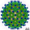
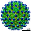
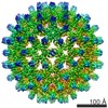
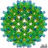
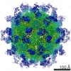
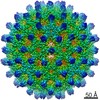
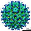
 Map data Map data | sub-tomogram averaging reconstruction of the hepatitis B capsid. | |||||||||
 Sample Sample |
| |||||||||
 Keywords Keywords | sub-tomogram averaging / RELION / maximum-likelihood / ultrastable gold substrates / hepatitis B capsid | |||||||||
| Function / homology |  Function and homology information Function and homology informationmicrotubule-dependent intracellular transport of viral material towards nucleus / T=4 icosahedral viral capsid / viral penetration into host nucleus / host cell / host cell cytoplasm / viral envelope / symbiont entry into host cell / structural molecule activity / DNA binding / RNA binding Similarity search - Function | |||||||||
| Biological species |   Hepatitis B virus Hepatitis B virus | |||||||||
| Method | subtomogram averaging / cryo EM / Resolution: 8.1 Å | |||||||||
 Authors Authors | Bharat TA / Russo CJ / Lowe J / Passmore LA / Scheres SHW | |||||||||
 Citation Citation | Journal: Structure / Year: 2015 Title: Advances in Single-Particle Electron Cryomicroscopy Structure Determination applied to Sub-tomogram Averaging. Authors: Tanmay A M Bharat / Christopher J Russo / Jan Löwe / Lori A Passmore / Sjors H W Scheres /  Abstract: Recent innovations in specimen preparation, data collection, and image processing have led to improved structure determination using single-particle electron cryomicroscopy (cryo-EM). Here we explore ...Recent innovations in specimen preparation, data collection, and image processing have led to improved structure determination using single-particle electron cryomicroscopy (cryo-EM). Here we explore some of these advances to improve structures determined using electron cryotomography (cryo-ET) and sub-tomogram averaging. We implement a new three-dimensional model for the contrast transfer function, and use this in a regularized likelihood optimization algorithm as implemented in the RELION program. Using direct electron detector data, we apply both single-particle analysis and sub-tomogram averaging to analyze radiation-induced movements of the specimen. As in single-particle cryo-EM, we find that significant sample movements occur during tomographic data acquisition, and that these movements are substantially reduced through the use of ultrastable gold substrates. We obtain a sub-nanometer resolution structure of the hepatitis B capsid, and show that reducing radiation-induced specimen movement may be central to attempts at further improving tomogram quality and resolution. | |||||||||
| History |
|
- Structure visualization
| Movie |
Movie viewer |
|---|---|
| Structure viewer | EM map: SurfViewMolmilJmol/JSmol |
| Supplemental images |

- Downloads & links
-EMDB archive
| Map data | emd_3015.map.gz | 9.1 MB | EMDB map data format | |
|---|---|---|---|---|
| Header (meta data) | emd-3015-v30.xmlemd-3015.xml | 8.9 KB 8.9 KB | Display Display | EMDB header |
| Images | 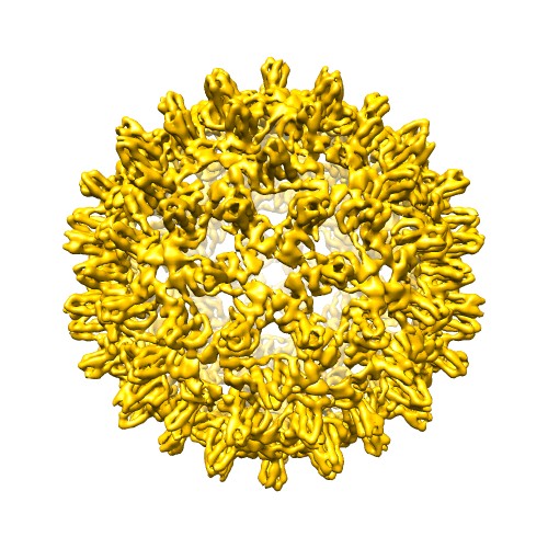 EMDB-3015-image500x500.jpg EMDB-3015-image500x500.jpg | 80.2 KB | ||
| Archive directory |  http://ftp.pdbj.org/pub/emdb/structures/EMD-3015ftp://ftp.pdbj.org/pub/emdb/structures/EMD-3015 http://ftp.pdbj.org/pub/emdb/structures/EMD-3015ftp://ftp.pdbj.org/pub/emdb/structures/EMD-3015 | HTTPS FTP |
-Related structure data
| Similar structure data |
|---|
-Links
| EMDB pages | EMDB (EBI/PDBe) / EMDataResource |
|---|---|
| Related items in Molecule of the Month |


-Map
| File | Download / File: emd_3015.map.gz / Format: CCP4 / Size: 51.5 MB / Type: IMAGE STORED AS FLOATING POINT NUMBER (4 BYTES) | ||||||||||||||||||||||||||||||||||||||||||||||||||||||||||||||||||||
|---|---|---|---|---|---|---|---|---|---|---|---|---|---|---|---|---|---|---|---|---|---|---|---|---|---|---|---|---|---|---|---|---|---|---|---|---|---|---|---|---|---|---|---|---|---|---|---|---|---|---|---|---|---|---|---|---|---|---|---|---|---|---|---|---|---|---|---|---|---|
| Annotation | sub-tomogram averaging reconstruction of the hepatitis B capsid. | ||||||||||||||||||||||||||||||||||||||||||||||||||||||||||||||||||||
| Projections & slices | Image control
Images are generated by Spider. | ||||||||||||||||||||||||||||||||||||||||||||||||||||||||||||||||||||
| Voxel size | X=Y=Z: 2.17 Å | ||||||||||||||||||||||||||||||||||||||||||||||||||||||||||||||||||||
| Density |
| ||||||||||||||||||||||||||||||||||||||||||||||||||||||||||||||||||||
| Symmetry | Space group: 1 | ||||||||||||||||||||||||||||||||||||||||||||||||||||||||||||||||||||
| Details | EMDB XML:
CCP4 map header:
| ||||||||||||||||||||||||||||||||||||||||||||||||||||||||||||||||||||
 Z (Sec.)
 Y (Row.)
 X (Col.)


-Supplemental data
- Sample components
-Entire : hepatitis B capsid
| Entire | Name: hepatitis B capsid |
|---|---|
| Components |
|
-Supramolecule #1000: hepatitis B capsid
| Supramolecule | Name: hepatitis B capsid / type: sample / ID: 1000 / Oligomeric state: icosahedral / Number unique components: 1 |
|---|---|
| Molecular weight | Theoretical: 3.8 MDa |
-Macromolecule #1: hepatitis B capsid
| Macromolecule | Name: hepatitis B capsid / type: protein_or_peptide / ID: 1 / Details: 10 nm gold particles were mixed with the sample. / Number of copies: 240 / Oligomeric state: icosahedral / Recombinant expression: Yes |
|---|---|
| Source (natural) | Organism: Hepatitis B virus |
| Molecular weight | Theoretical: 3.8 MDa |
| Recombinant expression | Organism:  |
| Sequence | UniProtKB: Capsid protein |
-Experimental details
-Structure determination
| Method | cryo EM |
|---|---|
 Processing Processing | subtomogram averaging |
| Aggregation state | particle |
-Sample preparation
| Grid | Details: 300 mesh ultrastable gold substrates with 1.2 micron holes (Russo and Passmore, Science 2014) |
|---|---|
| Vitrification | Cryogen name: ETHANE / Chamber humidity: 100 % / Instrument: FEI VITROBOT MARK IV |
- Electron microscopy
| Microscope | FEI TITAN KRIOS |
|---|---|
| Specialist optics | Energy filter - Name: Gatan Quantum Energy Filter / Energy filter - Lower energy threshold: 0.0 eV / Energy filter - Upper energy threshold: 20.0 eV |
| Details | Data was collected using SerialEM. |
| Date | Aug 28, 2014 |
| Image recording | Category: CCD / Film or detector model: GATAN K2 (4k x 4k) / Average electron dose: 60 e/Å2 |
| Electron beam | Acceleration voltage: 300 kV / Electron source:  FIELD EMISSION GUN FIELD EMISSION GUN |
| Electron optics | Illumination mode: FLOOD BEAM / Imaging mode: BRIGHT FIELD / Cs: 2.7 mm / Nominal defocus max: 5.6 µm / Nominal defocus min: 3.3 µm |
| Sample stage | Specimen holder model: FEI TITAN KRIOS AUTOGRID HOLDER / Tilt series - Axis1 - Min angle: -60 ° / Tilt series - Axis1 - Max angle: 60 ° |
| Experimental equipment |  Model: Titan Krios / Image courtesy: FEI Company |
-Image processing
| Details | Maximum-likelihood refinement was conducted in RELION using a weighted 3D CTF model. |
|---|---|
| Final reconstruction | Applied symmetry - Point group: I (icosahedral) / Algorithm: OTHER / Resolution.type: BY AUTHOR / Resolution: 8.1 Å / Resolution method: OTHER / Software - Name: IMOD, Tomo3D, RELION / Details: gold-standard FSC measurement was used. / Number subtomograms used: 1145 |
| CTF correction | Details: RELION 3D CTF Model |
