 Movie
Movie Controller

[English] 日本語
 Yorodumi
- EMDB-1169: ERj1p uses a universal ribosomal adaptor site to coordinate the 8... -
+ Open data

- Basic information
| Entry | Database: EMDB / ID: EMD-1169 | |||||||||
|---|---|---|---|---|---|---|---|---|---|---|
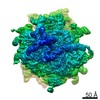
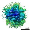
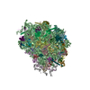
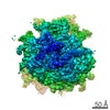
| Title | ERj1p uses a universal ribosomal adaptor site to coordinate the 80S ribosome at the membrane. | |||||||||
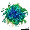
 Map data Map data | Volume of canine 80S+ErjC-dC | |||||||||
 Sample Sample |
| |||||||||
| Biological species |  | |||||||||
| Method | single particle reconstruction / cryo EM / Resolution: 20.0 Å | |||||||||
 Authors Authors | Blau M / Mullapudi S / Becker T / Dudek J / Zimmermann R / Penczek PA / Beckmann R | |||||||||
 Citation Citation | Journal: Nat Struct Mol Biol / Year: 2005 Title: ERj1p uses a universal ribosomal adaptor site to coordinate the 80S ribosome at the membrane. Authors: Michael Blau / Srinivas Mullapudi / Thomas Becker / Johanna Dudek / Richard Zimmermann / Pawel A Penczek / Roland Beckmann /  Abstract: Ribosomes translating secretory and membrane proteins are targeted to the endoplasmic reticulum membrane and attach to the protein-conducting channel and ribosome-associated membrane proteins (RAMPs). ...Ribosomes translating secretory and membrane proteins are targeted to the endoplasmic reticulum membrane and attach to the protein-conducting channel and ribosome-associated membrane proteins (RAMPs). Recently, a new RAMP, ERj1p, has been identified that recruits BiP to ribosomes and regulates translational activity. Here we present the cryo-EM structure of a ribosome-ERj1p complex, revealing how ERj1p coordinates the ribosome at the membrane and how allosteric effects may mediate ERj1p's regulatory activity. | |||||||||
| History |
|
- Structure visualization
| Movie |
 Movie viewer Movie viewer |
|---|---|
| Structure viewer | EM map: SurfViewMolmilJmol/JSmol |
| Supplemental images |

- Downloads & links
-EMDB archive
| Map data | emd_1169.map.gz | 10 MB | EMDB map data format | |
|---|---|---|---|---|
| Header (meta data) | emd-1169-v30.xmlemd-1169.xml | 6.7 KB 6.7 KB | Display Display | EMDB header |
| Images | 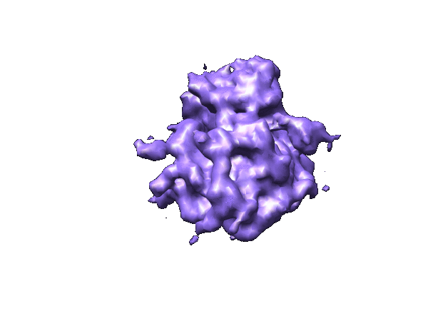 1169.gif 1169.gif | 43.5 KB | ||
| Archive directory |  http://ftp.pdbj.org/pub/emdb/structures/EMD-1169ftp://ftp.pdbj.org/pub/emdb/structures/EMD-1169 http://ftp.pdbj.org/pub/emdb/structures/EMD-1169ftp://ftp.pdbj.org/pub/emdb/structures/EMD-1169 | HTTPS FTP |
-Related structure data


-Links
| EMDB pages | EMDB (EBI/PDBe) / EMDataResource |
|---|---|
| Related items in Molecule of the Month |

-Map
| File | Download / File: emd_1169.map.gz / Format: CCP4 / Size: 11.1 MB / Type: IMAGE STORED AS FLOATING POINT NUMBER (4 BYTES) | ||||||||||||||||||||||||||||||||||||||||||||||||||||||||||||||||||||
|---|---|---|---|---|---|---|---|---|---|---|---|---|---|---|---|---|---|---|---|---|---|---|---|---|---|---|---|---|---|---|---|---|---|---|---|---|---|---|---|---|---|---|---|---|---|---|---|---|---|---|---|---|---|---|---|---|---|---|---|---|---|---|---|---|---|---|---|---|---|
| Annotation | Volume of canine 80S+ErjC-dC | ||||||||||||||||||||||||||||||||||||||||||||||||||||||||||||||||||||
| Projections & slices | Image control
Images are generated by Spider. | ||||||||||||||||||||||||||||||||||||||||||||||||||||||||||||||||||||
| Voxel size | X=Y=Z: 3.5 Å | ||||||||||||||||||||||||||||||||||||||||||||||||||||||||||||||||||||
| Density |
| ||||||||||||||||||||||||||||||||||||||||||||||||||||||||||||||||||||
| Symmetry | Space group: 1 | ||||||||||||||||||||||||||||||||||||||||||||||||||||||||||||||||||||
| Details | EMDB XML:
CCP4 map header:
| ||||||||||||||||||||||||||||||||||||||||||||||||||||||||||||||||||||
 Z (Sec.)
 Y (Row.)
 X (Col.)


-Supplemental data
- Sample components
-Entire : canine ribosome with bound ERj
| Entire | Name: canine ribosome with bound ERj |
|---|---|
| Components |
|
-Supramolecule #1000: canine ribosome with bound ERj
| Supramolecule | Name: canine ribosome with bound ERj / type: sample / ID: 1000 / Number unique components: 2 |
|---|
-Supramolecule #1: canine ribosome
| Supramolecule | Name: canine ribosome / type: complex / ID: 1 / Name.synonym: ribosome / Recombinant expression: No / Ribosome-details: ribosome-eukaryote: ALL |
|---|---|
| Source (natural) | Organism: |
-Macromolecule #1: ERjIp
| Macromolecule | Name: ERjIp / type: protein_or_peptide / ID: 1 / Name.synonym: ERjC-dC / Recombinant expression: Yes |
|---|---|
| Source (natural) | Organism: |
-Experimental details
-Structure determination
| Method | cryo EM |
|---|---|
 Processing Processing | single particle reconstruction |
| Aggregation state | particle |
-Sample preparation
| Vitrification | Cryogen name: ETHANE |
|---|
- Electron microscopy
| Microscope | JEOL KYOTO-3000SFF |
|---|---|
| Electron beam | Acceleration voltage: 300 kV / Electron source:  FIELD EMISSION GUN FIELD EMISSION GUN |
| Electron optics | Illumination mode: FLOOD BEAM / Imaging mode: BRIGHT FIELD |
| Sample stage | Specimen holder: Gathan / Specimen holder model: GATAN LIQUID NITROGEN |
-Image processing
| Final reconstruction | Applied symmetry - Point group: C1 (asymmetric) / Resolution.type: BY AUTHOR / Resolution: 20.0 Å |
|---|
