 ムービー
ムービー コントローラー


+ データを開く

- 基本情報
基本情報
| 登録情報 | データベース: EMDB / ID: EMD-1099 | |||||||||
|---|---|---|---|---|---|---|---|---|---|---|
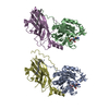
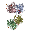
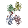
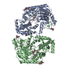
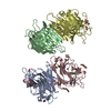
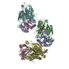
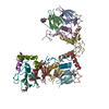
| タイトル | Architecture of CRM1/Exportin1 suggests how cooperativity is achieved during formation of a nuclear export complex. | |||||||||
 マップデータ マップデータ | 3D image reconstruction of human CRM1 | |||||||||
 試料 試料 |
| |||||||||
| 生物種 |  Homo sapiens (ヒト) Homo sapiens (ヒト) | |||||||||
| 手法 | 単粒子再構成法 / ネガティブ染色法 / 解像度: 22.0 Å | |||||||||
 データ登録者 データ登録者 | Petosa C | |||||||||
 引用 引用 | ジャーナル: Mol Cell / 年: 2004 タイトル: Architecture of CRM1/Exportin1 suggests how cooperativity is achieved during formation of a nuclear export complex. 著者: Carlo Petosa / Guy Schoehn / Peter Askjaer / Ulrike Bauer / Martine Moulin / Ulrich Steuerwald / Montserrat Soler-López / Florence Baudin / Iain W Mattaj / Christoph W Müller /  要旨: CRM1/Exportin1 mediates the nuclear export of proteins bearing a leucine-rich nuclear export signal (NES) by forming a cooperative ternary complex with the NES-bearing substrate and the small GTPase ...CRM1/Exportin1 mediates the nuclear export of proteins bearing a leucine-rich nuclear export signal (NES) by forming a cooperative ternary complex with the NES-bearing substrate and the small GTPase Ran. We present a structural model of human CRM1 based on a combination of X-ray crystallography, homology modeling, and electron microscopy. The architecture of CRM1 resembles that of the import receptor transportin1, with 19 HEAT repeats and a large loop implicated in Ran binding. Residues critical for NES recognition are identified adjacent to the cysteine residue targeted by leptomycin B (LMB), a specific CRM1 inhibitor. We present evidence that a conformational change of the Ran binding loop accounts for the cooperativity of Ran- and substrate binding and for the selective enhancement of CRM1-mediated export by the cofactor RanBP3. Our findings indicate that a single architectural and mechanistic framework can explain the divergent effects of RanGTP on substrate binding by many import and export receptors. | |||||||||
| 履歴 |
|
- 構造の表示
構造の表示
| ムービー |
 ムービービューア ムービービューア |
|---|---|
| 構造ビューア | EMマップ: SurfViewMolmilJmol/JSmol |
| 添付画像 |
 UCSF Chimera
- ダウンロードとリンク
-EMDBアーカイブ
| マップデータ | emd_1099.map.gz | 51.3 KB | EMDBマップデータ形式 | |
|---|---|---|---|---|
| ヘッダ (付随情報) | emd-1099-v30.xmlemd-1099.xml | 10.1 KB 10.1 KB | 表示 表示 | EMDBヘッダ |
| 画像 |  1099.gif 1099.gif | 11.3 KB | ||
| アーカイブディレクトリ |  http://ftp.pdbj.org/pub/emdb/structures/EMD-1099ftp://ftp.pdbj.org/pub/emdb/structures/EMD-1099 http://ftp.pdbj.org/pub/emdb/structures/EMD-1099ftp://ftp.pdbj.org/pub/emdb/structures/EMD-1099 | HTTPS FTP |
-検証レポート
| 文書・要旨 | emd_1099_validation.pdf.gz | 193.2 KB | 表示 | EMDB検証レポート |
|---|---|---|---|---|
| 文書・詳細版 | emd_1099_full_validation.pdf.gz | 192.4 KB | 表示 | |
| XML形式データ | emd_1099_validation.xml.gz | 4.7 KB | 表示 | |
| アーカイブディレクトリ | https://ftp.pdbj.org/pub/emdb/validation_reports/EMD-1099ftp://ftp.pdbj.org/pub/emdb/validation_reports/EMD-1099 | HTTPS FTP |
-関連構造データ


-リンク
| EMDBのページ | EMDB (EBI/PDBe) / EMDataResource |
|---|
-マップ
| ファイル | ダウンロード / ファイル: emd_1099.map.gz / 形式: CCP4 / 大きさ: 1001 KB / タイプ: IMAGE STORED AS FLOATING POINT NUMBER (4 BYTES) | ||||||||||||||||||||||||||||||||||||||||||||||||||||||||||||||||||||
|---|---|---|---|---|---|---|---|---|---|---|---|---|---|---|---|---|---|---|---|---|---|---|---|---|---|---|---|---|---|---|---|---|---|---|---|---|---|---|---|---|---|---|---|---|---|---|---|---|---|---|---|---|---|---|---|---|---|---|---|---|---|---|---|---|---|---|---|---|---|
| 注釈 | 3D image reconstruction of human CRM1 | ||||||||||||||||||||||||||||||||||||||||||||||||||||||||||||||||||||
| 投影像・断面図 | 画像のコントロール
画像は Spider により作成 | ||||||||||||||||||||||||||||||||||||||||||||||||||||||||||||||||||||
| ボクセルのサイズ | X=Y=Z: 3.5 Å | ||||||||||||||||||||||||||||||||||||||||||||||||||||||||||||||||||||
| 密度 |
| ||||||||||||||||||||||||||||||||||||||||||||||||||||||||||||||||||||
| 対称性 | 空間群: 1 | ||||||||||||||||||||||||||||||||||||||||||||||||||||||||||||||||||||
| 詳細 | EMDB XML:
CCP4マップ ヘッダ情報:
| ||||||||||||||||||||||||||||||||||||||||||||||||||||||||||||||||||||
 Z (Sec.)
 X (Row.)
 Y (Col.)


-添付データ
- 試料の構成要素
-全体 : human CRM1
| 全体 | 名称: human CRM1 |
|---|---|
| 要素 |
|
-超分子 #1000: human CRM1
| 超分子 | 名称: human CRM1 / タイプ: sample / ID: 1000 / 集合状態: monomer / Number unique components: 1 |
|---|---|
| 分子量 | 理論値: 123.385 KDa |
-分子 #1: CRM1
| 分子 | 名称: CRM1 / タイプ: protein_or_peptide / ID: 1 / コピー数: 1 / 集合状態: monomer / 組換発現: Yes |
|---|---|
| 由来(天然) | 生物種: Homo sapiens (ヒト) / 別称: human |
| 分子量 | 実験値: 123.385 KDa |
| 組換発現 | 生物種:  |
-実験情報
-構造解析
| 手法 | ネガティブ染色法 |
|---|---|
 解析 解析 | 単粒子再構成法 |
| 試料の集合状態 | particle |
-試料調製
| 濃度 | 0.1 mg/mL |
|---|---|
| 緩衝液 | pH: 7.5 / 詳細: 50 mM NaCl, 20 mM HEPES |
| 染色 | タイプ: NEGATIVE 詳細: Grids with adsorbed protein floated on 1% (w/v) sodium silicotungstate pH 7 |
| グリッド | 詳細: 400 mesh copper/ |
| 凍結 | 凍結剤: NONE |
- 電子顕微鏡法
| 顕微鏡 | JEOL 1200EXII |
|---|---|
| 温度 | 平均: 293 K |
| アライメント法 | Legacy - 非点収差: objective lens astigmatism was corrected at 200,000 times magnification |
| 詳細 | Low dose. Microscope JEOL 1200 EX II. |
| 撮影 | カテゴリ: FILM / フィルム・検出器のモデル: KODAK SO-163 FILM / デジタル化 - スキャナー: ZEISS SCAI / デジタル化 - サンプリング間隔: 14 µm / 実像数: 6 / 平均電子線量: 10 e/Å2 / Od range: 1 / ビット/ピクセル: 8 |
| Tilt angle min | 0 |
| Tilt angle max | 0 |
| 電子線 | 加速電圧: 100 kV / 電子線源: TUNGSTEN HAIRPIN |
| 電子光学系 | 倍率(補正後): 39950 / 照射モード: OTHER / 撮影モード: BRIGHT FIELD / Cs: 2.0 mm / 最大 デフォーカス(公称値): 2.0 µm / 最小 デフォーカス(公称値): 1.0 µm / 倍率(公称値): 40000 |
| 試料ステージ | 試料ホルダー: side entry room temperature holder / 試料ホルダーモデル: OTHER |
-画像解析
| CTF補正 | 詳細: Each particle |
|---|---|
| 最終 再構成 | 想定した対称性 - 点群: C1 (非対称) / アルゴリズム: OTHER / 解像度のタイプ: BY AUTHOR / 解像度: 22.0 Å / 解像度の算出法: FSC 0.5 CUT-OFF / ソフトウェア - 名称: SPIDER / 詳細: back projection of 196 class average using Spider / 使用した粒子像数: 5000 |
| 最終 角度割当 | 詳細: SPIDER : Theta : 180 degrees, phi 90 degrees |
| 最終 2次元分類 | クラス数: 196 |
-原子モデル構築 1
| 初期モデル | PDB ID: |
|---|---|
| ソフトウェア | 名称: Situs, Colores |
| 詳細 | Protocol: Rigid Body |
| 精密化 | 空間: REAL / プロトコル: RIGID BODY FIT |
