 Movie
Movie Controller

[English] 日本語
 Yorodumi
- EMDB-1099: Architecture of CRM1/Exportin1 suggests how cooperativity is achi... -
+ Open data

- Basic information
| Entry | Database: EMDB / ID: EMD-1099 | |||||||||
|---|---|---|---|---|---|---|---|---|---|---|
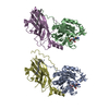
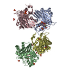
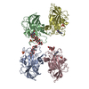
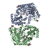
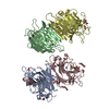
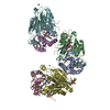
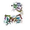
| Title | Architecture of CRM1/Exportin1 suggests how cooperativity is achieved during formation of a nuclear export complex. | |||||||||
 Map data Map data | 3D image reconstruction of human CRM1 | |||||||||
 Sample Sample |
| |||||||||
| Biological species |  Homo sapiens (human) Homo sapiens (human) | |||||||||
| Method | single particle reconstruction / negative staining / Resolution: 22.0 Å | |||||||||
 Authors Authors | Petosa C | |||||||||
 Citation Citation | Journal: Mol Cell / Year: 2004 Title: Architecture of CRM1/Exportin1 suggests how cooperativity is achieved during formation of a nuclear export complex. Authors: Carlo Petosa / Guy Schoehn / Peter Askjaer / Ulrike Bauer / Martine Moulin / Ulrich Steuerwald / Montserrat Soler-López / Florence Baudin / Iain W Mattaj / Christoph W Müller /  Abstract: CRM1/Exportin1 mediates the nuclear export of proteins bearing a leucine-rich nuclear export signal (NES) by forming a cooperative ternary complex with the NES-bearing substrate and the small GTPase ...CRM1/Exportin1 mediates the nuclear export of proteins bearing a leucine-rich nuclear export signal (NES) by forming a cooperative ternary complex with the NES-bearing substrate and the small GTPase Ran. We present a structural model of human CRM1 based on a combination of X-ray crystallography, homology modeling, and electron microscopy. The architecture of CRM1 resembles that of the import receptor transportin1, with 19 HEAT repeats and a large loop implicated in Ran binding. Residues critical for NES recognition are identified adjacent to the cysteine residue targeted by leptomycin B (LMB), a specific CRM1 inhibitor. We present evidence that a conformational change of the Ran binding loop accounts for the cooperativity of Ran- and substrate binding and for the selective enhancement of CRM1-mediated export by the cofactor RanBP3. Our findings indicate that a single architectural and mechanistic framework can explain the divergent effects of RanGTP on substrate binding by many import and export receptors. | |||||||||
| History |
|
- Structure visualization
| Movie |
 Movie viewer Movie viewer |
|---|---|
| Structure viewer | EM map: SurfViewMolmilJmol/JSmol |
| Supplemental images |
 UCSF Chimera
- Downloads & links
-EMDB archive
| Map data | emd_1099.map.gz | 51.3 KB | EMDB map data format | |
|---|---|---|---|---|
| Header (meta data) | emd-1099-v30.xmlemd-1099.xml | 10.1 KB 10.1 KB | Display Display | EMDB header |
| Images |  1099.gif 1099.gif | 11.3 KB | ||
| Archive directory |  http://ftp.pdbj.org/pub/emdb/structures/EMD-1099ftp://ftp.pdbj.org/pub/emdb/structures/EMD-1099 http://ftp.pdbj.org/pub/emdb/structures/EMD-1099ftp://ftp.pdbj.org/pub/emdb/structures/EMD-1099 | HTTPS FTP |
-Related structure data


-Links
| EMDB pages | EMDB (EBI/PDBe) / EMDataResource |
|---|
-Map
| File | Download / File: emd_1099.map.gz / Format: CCP4 / Size: 1001 KB / Type: IMAGE STORED AS FLOATING POINT NUMBER (4 BYTES) | ||||||||||||||||||||||||||||||||||||||||||||||||||||||||||||||||||||
|---|---|---|---|---|---|---|---|---|---|---|---|---|---|---|---|---|---|---|---|---|---|---|---|---|---|---|---|---|---|---|---|---|---|---|---|---|---|---|---|---|---|---|---|---|---|---|---|---|---|---|---|---|---|---|---|---|---|---|---|---|---|---|---|---|---|---|---|---|---|
| Annotation | 3D image reconstruction of human CRM1 | ||||||||||||||||||||||||||||||||||||||||||||||||||||||||||||||||||||
| Projections & slices | Image control
Images are generated by Spider. | ||||||||||||||||||||||||||||||||||||||||||||||||||||||||||||||||||||
| Voxel size | X=Y=Z: 3.5 Å | ||||||||||||||||||||||||||||||||||||||||||||||||||||||||||||||||||||
| Density |
| ||||||||||||||||||||||||||||||||||||||||||||||||||||||||||||||||||||
| Symmetry | Space group: 1 | ||||||||||||||||||||||||||||||||||||||||||||||||||||||||||||||||||||
| Details | EMDB XML:
CCP4 map header:
| ||||||||||||||||||||||||||||||||||||||||||||||||||||||||||||||||||||
 Z (Sec.)
 X (Row.)
 Y (Col.)


-Supplemental data
- Sample components
-Entire : human CRM1
| Entire | Name: human CRM1 |
|---|---|
| Components |
|
-Supramolecule #1000: human CRM1
| Supramolecule | Name: human CRM1 / type: sample / ID: 1000 / Oligomeric state: monomer / Number unique components: 1 |
|---|---|
| Molecular weight | Theoretical: 123.385 KDa |
-Macromolecule #1: CRM1
| Macromolecule | Name: CRM1 / type: protein_or_peptide / ID: 1 / Number of copies: 1 / Oligomeric state: monomer / Recombinant expression: Yes |
|---|---|
| Source (natural) | Organism: Homo sapiens (human) / synonym: human |
| Molecular weight | Experimental: 123.385 KDa |
| Recombinant expression | Organism:  |
-Experimental details
-Structure determination
| Method | negative staining |
|---|---|
 Processing Processing | single particle reconstruction |
| Aggregation state | particle |
-Sample preparation
| Concentration | 0.1 mg/mL |
|---|---|
| Buffer | pH: 7.5 / Details: 50 mM NaCl, 20 mM HEPES |
| Staining | Type: NEGATIVE Details: Grids with adsorbed protein floated on 1% (w/v) sodium silicotungstate pH 7 |
| Grid | Details: 400 mesh copper/ |
| Vitrification | Cryogen name: NONE |
- Electron microscopy
| Microscope | JEOL 1200EXII |
|---|---|
| Temperature | Average: 293 K |
| Alignment procedure | Legacy - Astigmatism: objective lens astigmatism was corrected at 200,000 times magnification |
| Details | Low dose. Microscope JEOL 1200 EX II. |
| Image recording | Category: FILM / Film or detector model: KODAK SO-163 FILM / Digitization - Scanner: ZEISS SCAI / Digitization - Sampling interval: 14 µm / Number real images: 6 / Average electron dose: 10 e/Å2 / Od range: 1 / Bits/pixel: 8 |
| Tilt angle min | 0 |
| Tilt angle max | 0 |
| Electron beam | Acceleration voltage: 100 kV / Electron source: TUNGSTEN HAIRPIN |
| Electron optics | Calibrated magnification: 39950 / Illumination mode: OTHER / Imaging mode: BRIGHT FIELD / Cs: 2.0 mm / Nominal defocus max: 2.0 µm / Nominal defocus min: 1.0 µm / Nominal magnification: 40000 |
| Sample stage | Specimen holder: side entry room temperature holder / Specimen holder model: OTHER |
-Image processing
| CTF correction | Details: Each particle |
|---|---|
| Final reconstruction | Applied symmetry - Point group: C1 (asymmetric) / Algorithm: OTHER / Resolution.type: BY AUTHOR / Resolution: 22.0 Å / Resolution method: FSC 0.5 CUT-OFF / Software - Name: SPIDER / Details: back projection of 196 class average using Spider / Number images used: 5000 |
| Final angle assignment | Details: SPIDER : Theta : 180 degrees, phi 90 degrees |
| Final two d classification | Number classes: 196 |
-Atomic model buiding 1
| Initial model | PDB ID: |
|---|---|
| Software | Name: Situs, Colores |
| Details | Protocol: Rigid Body |
| Refinement | Space: REAL / Protocol: RIGID BODY FIT |
