 Movie
Movie Controller

[English] 日本語
 Yorodumi
- EMDB-1094: Electron microscopic analysis of KvAP voltage-dependent K+ channe... -
+ Open data

- Basic information
| Entry | Database: EMDB / ID: EMD-1094 | |||||||||
|---|---|---|---|---|---|---|---|---|---|---|
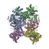
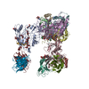
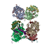
| Title | Electron microscopic analysis of KvAP voltage-dependent K+ channels in an open conformation. | |||||||||
 Map data Map data | A map of the KvAP/33H1Fab complex at 10.5 angstrom resolution. A threshold at 1.0 gives 340 kDa. | |||||||||
 Sample Sample |
| |||||||||
| Biological species |   Aeropyrum pernix (archaea) / Aeropyrum pernix (archaea) /  | |||||||||
| Method | single particle reconstruction / cryo EM / negative staining / Resolution: 10.5 Å | |||||||||
 Authors Authors | Jiang QX / Wang DN / MacKinnon R | |||||||||
 Citation Citation | Journal: Nature / Year: 2004 Title: Electron microscopic analysis of KvAP voltage-dependent K+ channels in an open conformation. Authors: Qiu-Xing Jiang / Da-Neng Wang / Roderick MacKinnon /  Abstract: Voltage-dependent ion channels serve as field-effect transistors by opening a gate in response to membrane voltage changes. The gate's response to voltage is mediated by voltage sensors, which are ...Voltage-dependent ion channels serve as field-effect transistors by opening a gate in response to membrane voltage changes. The gate's response to voltage is mediated by voltage sensors, which are arginine-containing structures that must move with respect to the membrane electric field. We have analysed by electron microscopy a voltage-dependent K(+) channel from Aeropyrum pernix (KvAP). Fab fragments were attached to 'voltage sensor paddles' and identified in the electron microscopy map at 10.5 A resolution. The extracellular surface location of the Fab fragments in the map is consistent with the membrane-depolarized, open conformation of the channel in electrophysiological experiments. Comparison of the map with a crystal structure demonstrates that the voltage sensor paddles are 'up' (that is, near the channel's extracellular surface) and situated at the protein-lipid interface. This finding supports the hypothesis that in response to changes in voltage the sensors move at the protein-lipid interface rather than in a gating pore surrounded by protein. | |||||||||
| History |
|
- Structure visualization
| Movie |
 Movie viewer Movie viewer |
|---|---|
| Structure viewer | EM map: SurfViewMolmilJmol/JSmol |
| Supplemental images |
- Downloads & links
-EMDB archive
| Map data | emd_1094.map.gz | 599.2 KB | EMDB map data format | |
|---|---|---|---|---|
| Header (meta data) | emd-1094-v30.xmlemd-1094.xml | 10.6 KB 10.6 KB | Display Display | EMDB header |
| Images |  1094.gif 1094.gif | 36.9 KB | ||
| Archive directory |  http://ftp.pdbj.org/pub/emdb/structures/EMD-1094ftp://ftp.pdbj.org/pub/emdb/structures/EMD-1094 http://ftp.pdbj.org/pub/emdb/structures/EMD-1094ftp://ftp.pdbj.org/pub/emdb/structures/EMD-1094 | HTTPS FTP |
-Related structure data
| Similar structure data |
|---|


-Links
| EMDB pages | EMDB (EBI/PDBe) / EMDataResource |
|---|
-Map
| File | Download / File: emd_1094.map.gz / Format: CCP4 / Size: 825.2 KB / Type: IMAGE STORED AS FLOATING POINT NUMBER (4 BYTES) | ||||||||||||||||||||||||||||||||||||||||||||||||||||||||||||||||||||
|---|---|---|---|---|---|---|---|---|---|---|---|---|---|---|---|---|---|---|---|---|---|---|---|---|---|---|---|---|---|---|---|---|---|---|---|---|---|---|---|---|---|---|---|---|---|---|---|---|---|---|---|---|---|---|---|---|---|---|---|---|---|---|---|---|---|---|---|---|---|
| Annotation | A map of the KvAP/33H1Fab complex at 10.5 angstrom resolution. A threshold at 1.0 gives 340 kDa. | ||||||||||||||||||||||||||||||||||||||||||||||||||||||||||||||||||||
| Projections & slices | Image control
Images are generated by Spider. | ||||||||||||||||||||||||||||||||||||||||||||||||||||||||||||||||||||
| Voxel size | X=Y=Z: 2.8 Å | ||||||||||||||||||||||||||||||||||||||||||||||||||||||||||||||||||||
| Density |
| ||||||||||||||||||||||||||||||||||||||||||||||||||||||||||||||||||||
| Symmetry | Space group: 1 | ||||||||||||||||||||||||||||||||||||||||||||||||||||||||||||||||||||
| Details | EMDB XML:
CCP4 map header:
| ||||||||||||||||||||||||||||||||||||||||||||||||||||||||||||||||||||
 Z (Sec.)
 Y (Row.)
 X (Col.)


-Supplemental data
- Sample components
-Entire : KvAP complexed with 33H1 Fab fragments
| Entire | Name: KvAP complexed with 33H1 Fab fragments |
|---|---|
| Components |
|
-Supramolecule #1000: KvAP complexed with 33H1 Fab fragments
| Supramolecule | Name: KvAP complexed with 33H1 Fab fragments / type: sample / ID: 1000 / Details: none / Oligomeric state: one / Number unique components: 2 |
|---|---|
| Molecular weight | Experimental: 340 KDa / Theoretical: 300 KDa Method: the sequence and the estimate of detergent micelle size |
-Macromolecule #1: KvAP potassium channel
| Macromolecule | Name: KvAP potassium channel / type: protein_or_peptide / ID: 1 / Name.synonym: KvAP / Details: one channel / Number of copies: 1 / Oligomeric state: tetramer / Recombinant expression: Yes |
|---|---|
| Source (natural) | Organism: Aeropyrum pernix (archaea) / synonym: A. pernix / Location in cell: plasma membrane |
| Molecular weight | Experimental: 120 KDa / Theoretical: 120 KDa |
| Recombinant expression | Organism:  |
-Macromolecule #2: 33H1 Fab fragments
| Macromolecule | Name: 33H1 Fab fragments / type: protein_or_peptide / ID: 2 / Name.synonym: 33H1Fab / Details: four Fab fragments / Number of copies: 4 / Oligomeric state: monomer / Recombinant expression: No |
|---|---|
| Source (natural) | Organism: |
| Molecular weight | Experimental: 180 KDa / Theoretical: 180 KDa |
-Experimental details
-Structure determination
| Method | negative staining, cryo EM |
|---|---|
 Processing Processing | single particle reconstruction |
| Aggregation state | particle |
-Sample preparation
| Concentration | 5.0 mg/mL |
|---|---|
| Buffer | pH: 7.4 Details: 40 mM KCl, 60 mM NaCl, 20 mM Tris-HCl pH 7.4, 5.0 mM DM |
| Staining | Type: NEGATIVE / Details: mix with equal volume of 65% ammonium molybdate |
| Grid | Details: 400 mesh grids with holey carbon film |
| Vitrification | Cryogen name: ETHANE / Chamber humidity: 60 % / Chamber temperature: 298 K / Instrument: HOMEMADE PLUNGER / Details: Vitrification instrument: home-made / Method: 8 |
- Electron microscopy
| Microscope | FEI/PHILIPS CM200T |
|---|---|
| Temperature | Average: 100 K |
| Alignment procedure | Legacy - Astigmatism: corrected at 200000 |
| Image recording | Category: FILM / Film or detector model: KODAK SO-163 FILM / Digitization - Scanner: ZEISS SCAI / Digitization - Sampling interval: 14 µm / Number real images: 37 / Average electron dose: 20 e/Å2 / Od range: 1.2 / Bits/pixel: 8 |
| Tilt angle min | 0 |
| Tilt angle max | 0 |
| Electron beam | Acceleration voltage: 200 kV / Electron source:  FIELD EMISSION GUN FIELD EMISSION GUN |
| Electron optics | Illumination mode: FLOOD BEAM / Imaging mode: BRIGHT FIELD / Cs: 2 mm / Nominal defocus max: 1.1 µm / Nominal defocus min: 0.5 µm / Nominal magnification: 50000 |
| Sample stage | Specimen holder: Oxford / Specimen holder model: OTHER |
-Image processing
| Details | the holes in the carbon film were covered with a thin-layer of carbon film. |
|---|---|
| CTF correction | Details: each particle |
| Final reconstruction | Applied symmetry - Point group: C4 (4 fold cyclic) / Algorithm: OTHER / Resolution.type: BY AUTHOR / Resolution: 10.5 Å / Resolution method: FSC 0.5 CUT-OFF / Software - Name: IMAGIC SPIDER / Number images used: 21379 |
| Final two d classification | Number classes: 496 |
