Japan Agency for Medical Research and Development (AMED)
JP18ek0109391
Japan
Japan Agency for Medical Research and Development (AMED)
JP18dm020719
Japan
National Institutes of Health/National Institute on Aging (NIH/NIA)
P30AG010133
United States
National Institutes of Health/National Institute of Neurological Disorders and Stroke (NIH/NINDS)
U01NS110437
United States
Citation
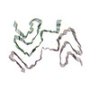
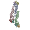
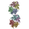
Journal: Nature / Year: 2020 Title: Structures of α-synuclein filaments from multiple system atrophy. Authors: Manuel Schweighauser / Yang Shi / Airi Tarutani / Fuyuki Kametani / Alexey G Murzin / Bernardino Ghetti / Tomoyasu Matsubara / Taisuke Tomita / Takashi Ando / Kazuko Hasegawa / Shigeo ...Authors: Manuel Schweighauser / Yang Shi / Airi Tarutani / Fuyuki Kametani / Alexey G Murzin / Bernardino Ghetti / Tomoyasu Matsubara / Taisuke Tomita / Takashi Ando / Kazuko Hasegawa / Shigeo Murayama / Mari Yoshida / Masato Hasegawa / Sjors H W Scheres / Michel Goedert / Abstract: Synucleinopathies, which include multiple system atrophy (MSA), Parkinson's disease, Parkinson's disease with dementia and dementia with Lewy bodies (DLB), are human neurodegenerative diseases. ...Synucleinopathies, which include multiple system atrophy (MSA), Parkinson's disease, Parkinson's disease with dementia and dementia with Lewy bodies (DLB), are human neurodegenerative diseases. Existing treatments are at best symptomatic. These diseases are characterized by the presence of, and believed to be caused by the formation of, filamentous inclusions of α-synuclein in brain cells. However, the structures of α-synuclein filaments from the human brain are unknown. Here, using cryo-electron microscopy, we show that α-synuclein inclusions from the brains of individuals with MSA are made of two types of filament, each of which consists of two different protofilaments. In each type of filament, non-proteinaceous molecules are present at the interface of the two protofilaments. Using two-dimensional class averaging, we show that α-synuclein filaments from the brains of individuals with MSA differ from those of individuals with DLB, which suggests that distinct conformers or strains characterize specific synucleinopathies. As is the case with tau assemblies, the structures of α-synuclein filaments extracted from the brains of individuals with MSA differ from those formed in vitro using recombinant proteins, which has implications for understanding the mechanisms of aggregate propagation and neurodegeneration in the human brain. These findings have diagnostic and potential therapeutic relevance, especially because of the unmet clinical need to be able to image filamentous α-synuclein inclusions in the human brain.
History
Deposition
Jan 30, 2020
-
Header (metadata) release
Feb 12, 2020
-
Map release
Feb 12, 2020
-
Update
Jul 2, 2025
-
Current status
Jul 2, 2025
Processing site: PDBe / Status: Released
-
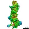
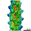
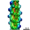
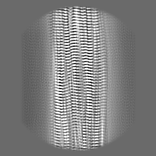
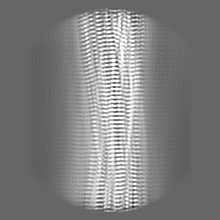
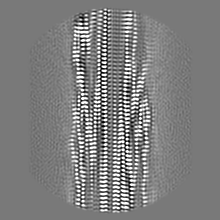
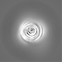
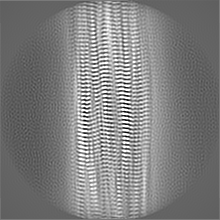
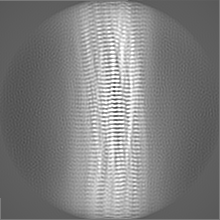
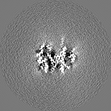
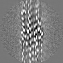
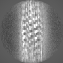
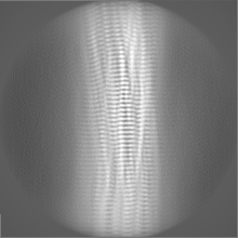
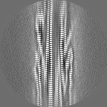
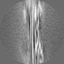
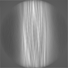
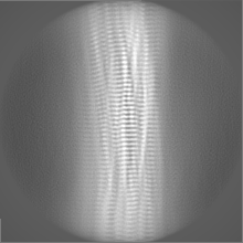
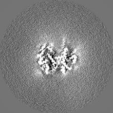
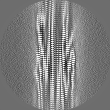
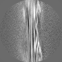
Structure visualization
Movie
Surface view with section colored by density value
EMPIAR-10358 (Title: CryoEM dataset of sarkosyl-insoluble fractions from the putamen of multiple system atrophy brain of case 2 Data size: 1.1 TB Data #1: Unaligned multi-frame movies [micrographs - multiframe] Data #2: Dose-weighted aligned micrographs [micrographs - single frame] Data #3: Polished particle stacks [picked particles - single frame - processed])
Film or detector model: GATAN K2 SUMMIT (4k x 4k) / Detector mode: COUNTING / Average electron dose: 47.5 e/Å2
Electron beam
Acceleration voltage: 300 kV / Electron source: FIELD EMISSION GUN
Electron optics
Illumination mode: FLOOD BEAM / Imaging mode: BRIGHT FIELD
Experimental equipment
Model: Titan Krios / Image courtesy: FEI Company
+
Image processing
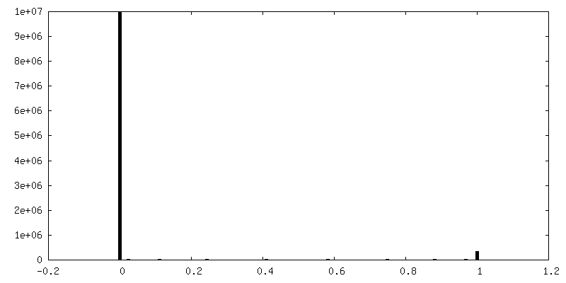
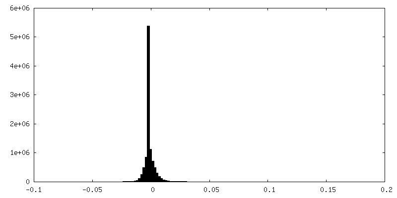
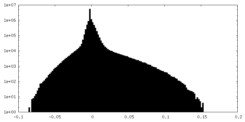
Final reconstruction
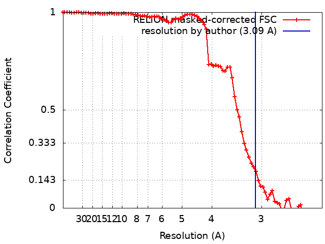
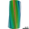
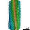
Applied symmetry - Helical parameters - Δz: 4.72 Å Applied symmetry - Helical parameters - Δ&Phi: -1.34 ° Applied symmetry - Helical parameters - Axial symmetry: C1 (asymmetric) Resolution.type: BY AUTHOR / Resolution: 3.09 Å / Resolution method: FSC 0.143 CUT-OFF Details: Helical reconstruction in RELION3. The initial model was reconstructed from reference-free 2-D class averages and the resolution of the cryo-EM map was good enough to do de-novo modelling. Number images used: 93137
CTF correction
Type: PHASE FLIPPING AND AMPLITUDE CORRECTION
Startup model
Type of model: INSILICO MODEL
Final angle assignment
Type: NOT APPLICABLE
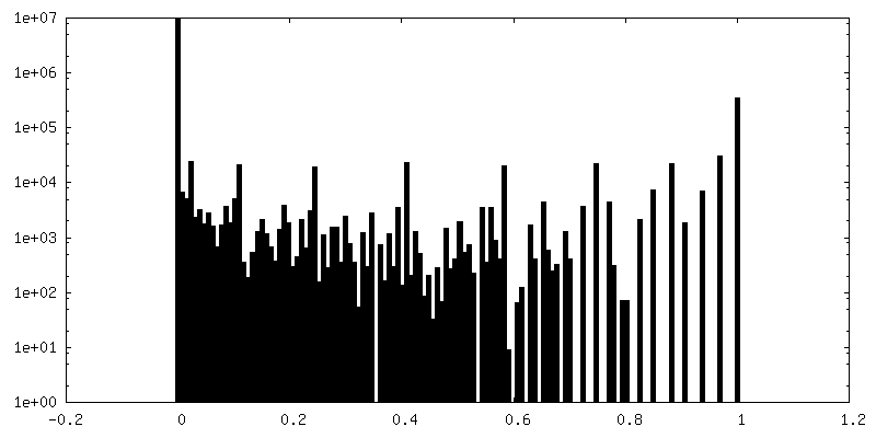
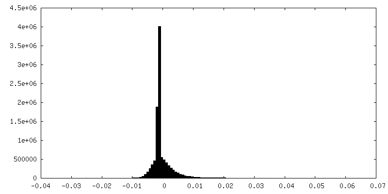
FSC plot (resolution estimation)
+
About Yorodumi
-
News
-
Feb 9, 2022. New format data for meta-information of EMDB entries
New format data for meta-information of EMDB entries
Version 3 of the EMDB header file is now the official format.
The previous official version 1.9 will be removed from the archive.
In the structure databanks used in Yorodumi, some data are registered as the other names, "COVID-19 virus" and "2019-nCoV". Here are the details of the virus and the list of structure data.
Jan 31, 2019. EMDB accession codes are about to change! (news from PDBe EMDB page)
EMDB accession codes are about to change! (news from PDBe EMDB page)
The allocation of 4 digits for EMDB accession codes will soon come to an end. Whilst these codes will remain in use, new EMDB accession codes will include an additional digit and will expand incrementally as the available range of codes is exhausted. The current 4-digit format prefixed with “EMD-” (i.e. EMD-XXXX) will advance to a 5-digit format (i.e. EMD-XXXXX), and so on. It is currently estimated that the 4-digit codes will be depleted around Spring 2019, at which point the 5-digit format will come into force.
The EM Navigator/Yorodumi systems omit the EMD- prefix.
Related info.:Q: What is EMD? / ID/Accession-code notation in Yorodumi/EM Navigator
Yorodumi is a browser for structure data from EMDB, PDB, SASBDB, etc.
This page is also the successor to EM Navigator detail page, and also detail information page/front-end page for Omokage search.
The word "yorodu" (or yorozu) is an old Japanese word meaning "ten thousand". "mi" (miru) is to see.
Related info.:EMDB / PDB / SASBDB / Comparison of 3 databanks / Yorodumi Search / Aug 31, 2016. New EM Navigator & Yorodumi / Yorodumi Papers / Jmol/JSmol / Function and homology information / Changes in new EM Navigator and Yorodumi
 Movie
Movie Controller


 Open data
 Basic information
 Map data
 Sample
 Keywords
 Function and homology information
 Homo sapiens (human)
 Authors
 United Kingdom,
  Japan,
  United States, 7 items
  Citation
 Structure visualization
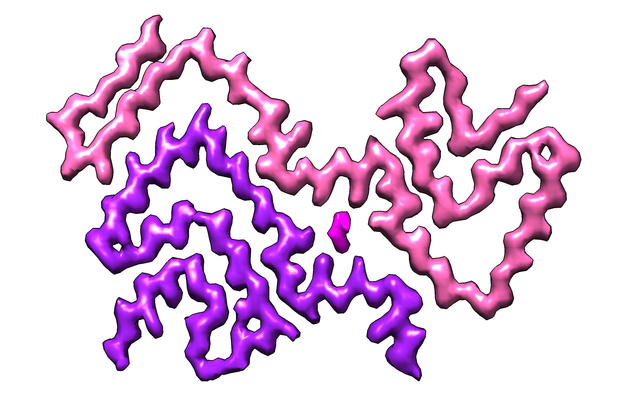
 Downloads & links
 emd_10652.png
 http://ftp.pdbj.org/pub/emdb/structures/EMD-10652


 Z (Sec.)
 Y (Row.)
 X (Col.)


 Sample components
 Processing
 Electron microscopy
 FIELD EMISSION GUN