 Movie
Movie Controller


+ Open data

- Basic information
| Entry | Database: PDB / ID: 6xyp | |||||||||||||||||||||||||||||||||||||||||||||||||||||||||||||||||||||||||||||||||||||||||||||
|---|---|---|---|---|---|---|---|---|---|---|---|---|---|---|---|---|---|---|---|---|---|---|---|---|---|---|---|---|---|---|---|---|---|---|---|---|---|---|---|---|---|---|---|---|---|---|---|---|---|---|---|---|---|---|---|---|---|---|---|---|---|---|---|---|---|---|---|---|---|---|---|---|---|---|---|---|---|---|---|---|---|---|---|---|---|---|---|---|---|---|---|---|---|---|
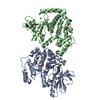
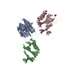
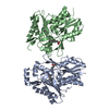
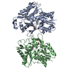
| Title | Multiple system atrophy Type II-1 alpha-synuclein filament | |||||||||||||||||||||||||||||||||||||||||||||||||||||||||||||||||||||||||||||||||||||||||||||
 Components Components | Alpha-synuclein | |||||||||||||||||||||||||||||||||||||||||||||||||||||||||||||||||||||||||||||||||||||||||||||
 Keywords Keywords | PROTEIN FIBRIL / multiple system atrophy / alpha-synuclein filament | |||||||||||||||||||||||||||||||||||||||||||||||||||||||||||||||||||||||||||||||||||||||||||||
| Function / homology |  Function and homology information Function and homology informationnegative regulation of mitochondrial electron transport, NADH to ubiquinone / negative regulation of dopamine uptake involved in synaptic transmission / negative regulation of norepinephrine uptake / response to desipramine / positive regulation of SNARE complex assembly / positive regulation of hydrogen peroxide catabolic process / supramolecular fiber / negative regulation of chaperone-mediated autophagy / regulation of synaptic vesicle recycling / regulation of reactive oxygen species biosynthetic process ...negative regulation of mitochondrial electron transport, NADH to ubiquinone / negative regulation of dopamine uptake involved in synaptic transmission / negative regulation of norepinephrine uptake / response to desipramine / positive regulation of SNARE complex assembly / positive regulation of hydrogen peroxide catabolic process / supramolecular fiber / negative regulation of chaperone-mediated autophagy / regulation of synaptic vesicle recycling / regulation of reactive oxygen species biosynthetic process / positive regulation of protein localization to cell periphery / negative regulation of exocytosis / dopamine biosynthetic process / dopamine uptake involved in synaptic transmission / negative regulation of dopamine metabolic process / response to iron(II) ion / negative regulation of platelet-derived growth factor receptor signaling pathway / SNARE complex assembly / negative regulation of thrombin-activated receptor signaling pathway / negative regulation of microtubule polymerization / synaptic vesicle priming / synaptic vesicle transport / Lewy body / synaptic vesicle exocytosis / regulation of norepinephrine uptake / transporter regulator activity / protein kinase inhibitor activity / positive regulation of inositol phosphate biosynthetic process / positive regulation of receptor recycling / regulation of dopamine secretion / cuprous ion binding / positive regulation of exocytosis / nuclear outer membrane / dynein complex binding / synaptic transmission, dopaminergic / response to magnesium ion / positive regulation of endocytosis / negative regulation of serotonin uptake / cysteine-type endopeptidase inhibitor activity / kinesin binding / regulation of presynapse assembly / alpha-tubulin binding / synaptic vesicle endocytosis / beta-tubulin binding / phospholipase binding / behavioral response to cocaine / cellular response to fibroblast growth factor stimulus / supramolecular fiber organization / response to type II interferon / cellular response to epinephrine stimulus / inclusion body / response to interleukin-1 / Hsp70 protein binding / positive regulation of release of sequestered calcium ion into cytosol / cellular response to copper ion / axon terminus / enzyme inhibitor activity / glutathione metabolic process / SNARE binding / protein tetramerization / regulation of microtubule cytoskeleton organization / receptor internalization / phosphoprotein binding / microglial cell activation / protein destabilization / protein sequestering activity / tubulin binding / ferrous iron binding / phospholipid binding / synapse organization / PKR-mediated signaling / tau protein binding / enzyme activator activity / positive regulation of inflammatory response / terminal bouton / actin cytoskeleton / synaptic vesicle membrane / negative regulation of neuron apoptotic process / response to lipopolysaccharide / growth cone / actin binding / cellular response to oxidative stress / histone binding / cell cortex / microtubule binding / amyloid fibril formation / mitochondrial outer membrane / lysosome / oxidoreductase activity / transcription cis-regulatory region binding / mitochondrial inner membrane / positive regulation of apoptotic process / ribosome / mitochondrial matrix / Amyloid fiber formation / copper ion binding / protein domain specific binding / axon / neuronal cell body / calcium ion binding Similarity search - Function | |||||||||||||||||||||||||||||||||||||||||||||||||||||||||||||||||||||||||||||||||||||||||||||
| Biological species |  Homo sapiens (human) Homo sapiens (human) | |||||||||||||||||||||||||||||||||||||||||||||||||||||||||||||||||||||||||||||||||||||||||||||
| Method | ELECTRON MICROSCOPY / helical reconstruction / cryo EM / Resolution: 3.29 Å | |||||||||||||||||||||||||||||||||||||||||||||||||||||||||||||||||||||||||||||||||||||||||||||
 Authors Authors | Schweighauser, M. / Shi, Y. / Tarutani, A. / Kametani, F. / Murzin, A.G. / Ghetti, B. / Matsubara, T. / Tomita, T. / Ando, T. / Hasegawa, K. ...Schweighauser, M. / Shi, Y. / Tarutani, A. / Kametani, F. / Murzin, A.G. / Ghetti, B. / Matsubara, T. / Tomita, T. / Ando, T. / Hasegawa, K. / Murayama, S. / Yoshida, M. / Hasegawa, M. / Scheres, S.H.W. / Goedert, M. | |||||||||||||||||||||||||||||||||||||||||||||||||||||||||||||||||||||||||||||||||||||||||||||
| Funding support |  United Kingdom, United Kingdom,  Japan, Japan,  United States, 7items United States, 7items
| |||||||||||||||||||||||||||||||||||||||||||||||||||||||||||||||||||||||||||||||||||||||||||||
 Citation Citation | Journal: Nature / Year: 2020 Title: Structures of α-synuclein filaments from multiple system atrophy. Authors: Manuel Schweighauser / Yang Shi / Airi Tarutani / Fuyuki Kametani / Alexey G Murzin / Bernardino Ghetti / Tomoyasu Matsubara / Taisuke Tomita / Takashi Ando / Kazuko Hasegawa / Shigeo ...Authors: Manuel Schweighauser / Yang Shi / Airi Tarutani / Fuyuki Kametani / Alexey G Murzin / Bernardino Ghetti / Tomoyasu Matsubara / Taisuke Tomita / Takashi Ando / Kazuko Hasegawa / Shigeo Murayama / Mari Yoshida / Masato Hasegawa / Sjors H W Scheres / Michel Goedert / Abstract: Synucleinopathies, which include multiple system atrophy (MSA), Parkinson's disease, Parkinson's disease with dementia and dementia with Lewy bodies (DLB), are human neurodegenerative diseases. ...Synucleinopathies, which include multiple system atrophy (MSA), Parkinson's disease, Parkinson's disease with dementia and dementia with Lewy bodies (DLB), are human neurodegenerative diseases. Existing treatments are at best symptomatic. These diseases are characterized by the presence of, and believed to be caused by the formation of, filamentous inclusions of α-synuclein in brain cells. However, the structures of α-synuclein filaments from the human brain are unknown. Here, using cryo-electron microscopy, we show that α-synuclein inclusions from the brains of individuals with MSA are made of two types of filament, each of which consists of two different protofilaments. In each type of filament, non-proteinaceous molecules are present at the interface of the two protofilaments. Using two-dimensional class averaging, we show that α-synuclein filaments from the brains of individuals with MSA differ from those of individuals with DLB, which suggests that distinct conformers or strains characterize specific synucleinopathies. As is the case with tau assemblies, the structures of α-synuclein filaments extracted from the brains of individuals with MSA differ from those formed in vitro using recombinant proteins, which has implications for understanding the mechanisms of aggregate propagation and neurodegeneration in the human brain. These findings have diagnostic and potential therapeutic relevance, especially because of the unmet clinical need to be able to image filamentous α-synuclein inclusions in the human brain. | |||||||||||||||||||||||||||||||||||||||||||||||||||||||||||||||||||||||||||||||||||||||||||||
| History |
|
- Structure visualization
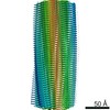
| Movie |
Movie viewer |
|---|---|
| Structure viewer | Molecule: MolmilJmol/JSmol |
- Downloads & links
-Download
| PDBx/mmCIF format | 6xyp.cif.gz | 123.4 KB | Display | PDBx/mmCIF format |
|---|---|---|---|---|
| PDB format | pdb6xyp.ent.gz | 94.3 KB | Display | PDB format |
| PDBx/mmJSON format | 6xyp.json.gz | Tree view | PDBx/mmJSON format | |
| Others |  Other downloads Other downloads |
-Validation report
| Arichive directory | https://data.pdbj.org/pub/pdb/validation_reports/xy/6xypftp://data.pdbj.org/pub/pdb/validation_reports/xy/6xyp | HTTPS FTP |
|---|
-Related structure data
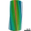
| Related structure data | 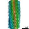 10651MC 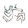 6xyoC 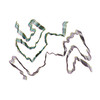 6xyqC M: map data used to model this data C: citing same article ( |
|---|---|
| Similar structure data | |
| EM raw data | EMPIAR-10358 (Title: CryoEM dataset of sarkosyl-insoluble fractions from the putamen of multiple system atrophy brain of case 2 Data size: 1.1 TB Data #1: Unaligned multi-frame movies [micrographs - multiframe] Data #2: Dose-weighted aligned micrographs [micrographs - single frame] Data #3: Polished particle stacks [picked particles - single frame - processed]) |
-Links
 PDBj
PDBj
- Assembly
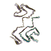
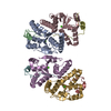
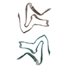
| Deposited unit | 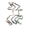
|
|---|---|
| 1 |
|
-Components
| #1: Protein | Mass: 14476.108 Da / Num. of mol.: 10 / Source method: isolated from a natural source / Source: (natural) Homo sapiens (human) / References: UniProt: P37840Has protein modification | N | |
|---|
-Experimental details
-Experiment
| Experiment | Method: ELECTRON MICROSCOPY |
|---|---|
| EM experiment | Aggregation state: FILAMENT / 3D reconstruction method: helical reconstruction |
- Sample preparation
| Component | Name: Sarkosyl-insoluble fractions from the putamen of multiple system atrophy brain Type: TISSUE / Entity ID: all / Source: NATURAL |
|---|---|
| Source (natural) | Organism: Homo sapiens (human) |
| Buffer solution | pH: 7.4 |
| Specimen | Embedding applied: NO / Shadowing applied: NO / Staining applied: NO / Vitrification applied: YES |
| Vitrification | Cryogen name: ETHANE |
- Electron microscopy imaging
| Experimental equipment |  Model: Titan Krios / Image courtesy: FEI Company |
|---|---|
| Microscopy | Model: FEI TITAN KRIOS |
| Electron gun | Electron source:  FIELD EMISSION GUN / Accelerating voltage: 300 kV / Illumination mode: FLOOD BEAM FIELD EMISSION GUN / Accelerating voltage: 300 kV / Illumination mode: FLOOD BEAM |
| Electron lens | Mode: BRIGHT FIELD |
| Image recording | Electron dose: 47.5 e/Å2 / Detector mode: COUNTING / Film or detector model: GATAN K2 SUMMIT (4k x 4k) |
- Processing
| Software | Name: PHENIX / Version: dev_3580: / Classification: refinement | ||||||||||||||||||||||||
|---|---|---|---|---|---|---|---|---|---|---|---|---|---|---|---|---|---|---|---|---|---|---|---|---|---|
| EM software | Name: PHENIX / Category: model refinement | ||||||||||||||||||||||||
| CTF correction | Type: PHASE FLIPPING AND AMPLITUDE CORRECTION | ||||||||||||||||||||||||
| Helical symmerty | Angular rotation/subunit: -1.41 ° / Axial rise/subunit: 4.72 Å / Axial symmetry: C1 | ||||||||||||||||||||||||
| 3D reconstruction | Resolution: 3.29 Å / Resolution method: FSC 0.143 CUT-OFF / Num. of particles: 23983 Details: Helical reconstruction in RELION3. The initial model was reconstructed from reference-free 2-D class averages and the resolution of the cryo-EM map was good enough to do de-novo modelling. Symmetry type: HELICAL | ||||||||||||||||||||||||
| Refine LS restraints |
|
