- EMDB-10513: Structure of the NDH-1MS complex from Thermosynechococcus elongatus -
+
Open data
ID or keywords:
Loading...
-
Basic information
Entry
Database: EMDB / ID: EMD-10513
Title
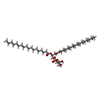
Structure of the NDH-1MS complex from Thermosynechococcus elongatus
Map data
Structure of the NDH-1MS complex from Thermosynechococcus elongatus
Sample
Complex: NDH-1MS
Protein or peptide: x 18 types
Ligand: x 8 types
Keywords
carbon concentrating photosynthetic complex I / proton pump / MEMBRANE PROTEIN
Function / homology
Function and homology information
Translocases; Catalysing the translocation of protons; Linked to oxidoreductase reactions / NADH dehydrogenase complex / transmembrane transporter complex / photosynthetic electron transport chain / oxidoreductase activity, acting on NAD(P)H, quinone or similar compound as acceptor / plasma membrane-derived thylakoid membrane / photosynthesis, light reaction / ubiquinone binding / electron transport coupled proton transport / NADH dehydrogenase activity ...Translocases; Catalysing the translocation of protons; Linked to oxidoreductase reactions / NADH dehydrogenase complex / transmembrane transporter complex / photosynthetic electron transport chain / oxidoreductase activity, acting on NAD(P)H, quinone or similar compound as acceptor / plasma membrane-derived thylakoid membrane / photosynthesis, light reaction / ubiquinone binding / electron transport coupled proton transport / NADH dehydrogenase activity / respiratory chain complex I / NADH dehydrogenase (ubiquinone) activity / quinone binding / ATP synthesis coupled electron transport / endomembrane system / aerobic respiration / NAD binding / 4 iron, 4 sulfur cluster binding / iron ion binding / : / membrane / plasma membrane Similarity search - Function
Journal: Nat Commun / Year: 2020 Title: Redox-coupled proton pumping drives carbon concentration in the photosynthetic complex I. Authors: Jan M Schuller / Patricia Saura / Jacqueline Thiemann / Sandra K Schuller / Ana P Gamiz-Hernandez / Genji Kurisu / Marc M Nowaczyk / Ville R I Kaila / Abstract: Photosynthetic organisms capture light energy to drive their energy metabolism, and employ the chemical reducing power to convert carbon dioxide (CO) into organic molecules. Photorespiration, ...Photosynthetic organisms capture light energy to drive their energy metabolism, and employ the chemical reducing power to convert carbon dioxide (CO) into organic molecules. Photorespiration, however, significantly reduces the photosynthetic yields. To survive under low CO concentrations, cyanobacteria evolved unique carbon-concentration mechanisms that enhance the efficiency of photosynthetic CO fixation, for which the molecular principles have remained unknown. We show here how modular adaptations enabled the cyanobacterial photosynthetic complex I to concentrate CO using a redox-driven proton-pumping machinery. Our cryo-electron microscopy structure at 3.2 Å resolution shows a catalytic carbonic anhydrase module that harbours a Zn active site, with connectivity to proton-pumping subunits that are activated by electron transfer from photosystem I. Our findings illustrate molecular principles in the photosynthetic complex I machinery that enabled cyanobacteria to survive in drastically changing CO conditions.
History
Deposition
Nov 27, 2019
-
Header (metadata) release
Feb 19, 2020
-
Map release
Feb 19, 2020
-
Update
Oct 1, 2025
-
Current status
Oct 1, 2025
Processing site: PDBe / Status: Released
-
Structure visualization
Movie
Surface view with section colored by density value
In the structure databanks used in Yorodumi, some data are registered as the other names, "COVID-19 virus" and "2019-nCoV". Here are the details of the virus and the list of structure data.
Jan 31, 2019. EMDB accession codes are about to change! (news from PDBe EMDB page)
EMDB accession codes are about to change! (news from PDBe EMDB page)
The allocation of 4 digits for EMDB accession codes will soon come to an end. Whilst these codes will remain in use, new EMDB accession codes will include an additional digit and will expand incrementally as the available range of codes is exhausted. The current 4-digit format prefixed with “EMD-” (i.e. EMD-XXXX) will advance to a 5-digit format (i.e. EMD-XXXXX), and so on. It is currently estimated that the 4-digit codes will be depleted around Spring 2019, at which point the 5-digit format will come into force.
The EM Navigator/Yorodumi systems omit the EMD- prefix.
Related info.:Q: What is EMD? / ID/Accession-code notation in Yorodumi/EM Navigator
Yorodumi is a browser for structure data from EMDB, PDB, SASBDB, etc.
This page is also the successor to EM Navigator detail page, and also detail information page/front-end page for Omokage search.
The word "yorodu" (or yorozu) is an old Japanese word meaning "ten thousand". "mi" (miru) is to see.
Related info.:EMDB / PDB / SASBDB / Comparison of 3 databanks / Yorodumi Search / Aug 31, 2016. New EM Navigator & Yorodumi / Yorodumi Papers / Jmol/JSmol / Function and homology information / Changes in new EM Navigator and Yorodumi
 Movie
Movie Controller

 Yorodumi
 Open data
 Basic information
 Map data
 Sample
 Keywords
 Function and homology information
 Thermosynechococcus elongatus BP-1 (bacteria) /
  Authors
 Germany, 4 items
  Citation

 Structure visualization
 Downloads & links
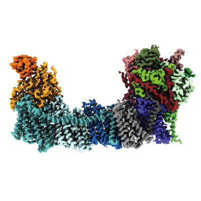 emd_10513.png
 http://ftp.pdbj.org/pub/emdb/structures/EMD-10513


 Z (Sec.)
 Y (Row.)
 X (Col.)


 Sample components


 Processing
 Electron microscopy
 FIELD EMISSION GUN
