 Movie
Movie Controller


+ Open data

- Basic information
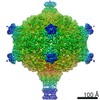
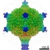
| Entry | Database: EMDB / ID: EMD-10442 | ||||||||||||||||||||||||
|---|---|---|---|---|---|---|---|---|---|---|---|---|---|---|---|---|---|---|---|---|---|---|---|---|---|
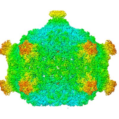
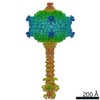
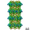
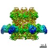
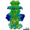
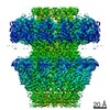
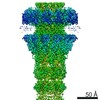
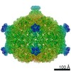
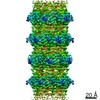
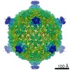
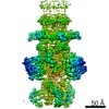
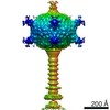
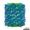
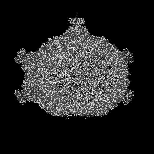
| Title | Capsid of native GTA particle computed with C5 symmetry | ||||||||||||||||||||||||
 Map data Map data | capsid of native GTA particle, C5 symmetrized | ||||||||||||||||||||||||
 Sample Sample |
| ||||||||||||||||||||||||
 Keywords Keywords | "capsid" / "gene transfer agent" / "bacteriophage" / "HK97" / VIRUS | ||||||||||||||||||||||||
| Function / homology | : / Phage capsid / Phage capsid family / : / : / Phage major capsid protein, HK97 family Function and homology information Function and homology information | ||||||||||||||||||||||||
| Biological species |  Rhodobacter capsulatus (bacteria) Rhodobacter capsulatus (bacteria) | ||||||||||||||||||||||||
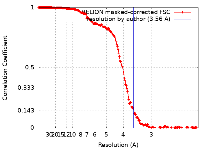
| Method | single particle reconstruction / cryo EM / Resolution: 3.56 Å | ||||||||||||||||||||||||
 Authors Authors | Bardy P / Fuzik T | ||||||||||||||||||||||||
| Funding support |  Czech Republic, 7 items Czech Republic, 7 items
| ||||||||||||||||||||||||
 Citation Citation | Journal: Nat Commun / Year: 2020 Title: Structure and mechanism of DNA delivery of a gene transfer agent. Authors: Pavol Bárdy / Tibor Füzik / Dominik Hrebík / Roman Pantůček / J Thomas Beatty / Pavel Plevka /  Abstract: Alphaproteobacteria, which are the most abundant microorganisms of temperate oceans, produce phage-like particles called gene transfer agents (GTAs) that mediate lateral gene exchange. However, the ...Alphaproteobacteria, which are the most abundant microorganisms of temperate oceans, produce phage-like particles called gene transfer agents (GTAs) that mediate lateral gene exchange. However, the mechanism by which GTAs deliver DNA into cells is unknown. Here we present the structure of the GTA of Rhodobacter capsulatus (RcGTA) and describe the conformational changes required for its DNA ejection. The structure of RcGTA resembles that of a tailed phage, but it has an oblate head shortened in the direction of the tail axis, which limits its packaging capacity to less than 4,500 base pairs of linear double-stranded DNA. The tail channel of RcGTA contains a trimer of proteins that possess features of both tape measure proteins of long-tailed phages from the family Siphoviridae and tail needle proteins of short-tailed phages from the family Podoviridae. The opening of a constriction within the RcGTA baseplate enables the ejection of DNA into bacterial periplasm. | ||||||||||||||||||||||||
| History |
|
- Structure visualization
| Movie |
Movie viewer |
|---|---|
| Structure viewer | EM map: SurfViewMolmilJmol/JSmol |
| Supplemental images |
- Downloads & links
-EMDB archive
| Map data | emd_10442.map.gz | 76.9 MB | EMDB map data format | |
|---|---|---|---|---|
| Header (meta data) | emd-10442-v30.xmlemd-10442.xml | 19.4 KB 19.4 KB | Display Display | EMDB header |
| FSC (resolution estimation) | emd_10442_fsc.xml | 18.1 KB | Display | FSC data file |
| Images |  emd_10442.png emd_10442.png | 239.3 KB | ||
| Filedesc metadata | emd-10442.cif.gz | 6.6 KB | ||
| Archive directory |  http://ftp.pdbj.org/pub/emdb/structures/EMD-10442ftp://ftp.pdbj.org/pub/emdb/structures/EMD-10442 http://ftp.pdbj.org/pub/emdb/structures/EMD-10442ftp://ftp.pdbj.org/pub/emdb/structures/EMD-10442 | HTTPS FTP |
-Related structure data
| Related structure data | 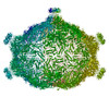 6tb9MC 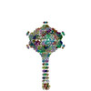 6tbaC 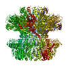 6te8C 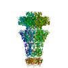 6te9C  6teaC  6tebC 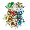 6tehC 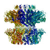 6to8C 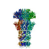 6toaC  6tsuC 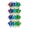 6tsvC 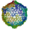 6tswC 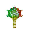 6tuiC C: citing same article ( M: atomic model generated by this map |
|---|---|
| Similar structure data |
-Links
| EMDB pages | EMDB (EBI/PDBe) / EMDataResource |
|---|---|
| Related items in Molecule of the Month |


-Map
| File | Download / File: emd_10442.map.gz / Format: CCP4 / Size: 512 MB / Type: IMAGE STORED AS FLOATING POINT NUMBER (4 BYTES) | ||||||||||||||||||||||||||||||||||||||||||||||||||||||||||||
|---|---|---|---|---|---|---|---|---|---|---|---|---|---|---|---|---|---|---|---|---|---|---|---|---|---|---|---|---|---|---|---|---|---|---|---|---|---|---|---|---|---|---|---|---|---|---|---|---|---|---|---|---|---|---|---|---|---|---|---|---|---|
| Annotation | capsid of native GTA particle, C5 symmetrized | ||||||||||||||||||||||||||||||||||||||||||||||||||||||||||||
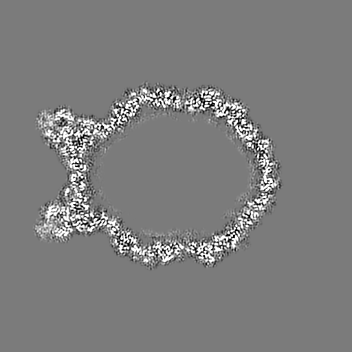
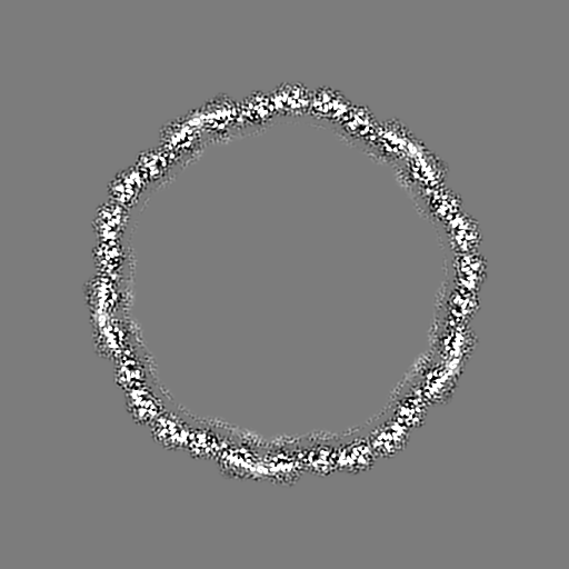
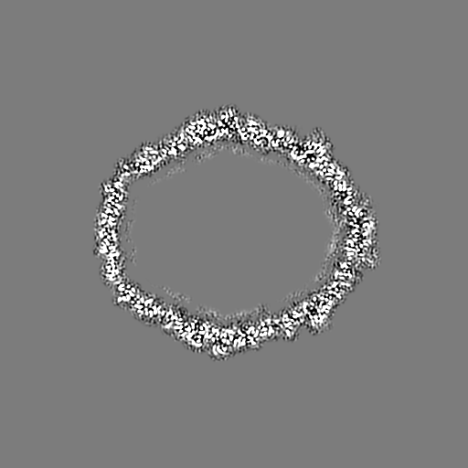
| Projections & slices | Image control
Images are generated by Spider. | ||||||||||||||||||||||||||||||||||||||||||||||||||||||||||||
| Voxel size | X=Y=Z: 1.063 Å | ||||||||||||||||||||||||||||||||||||||||||||||||||||||||||||
| Density |
| ||||||||||||||||||||||||||||||||||||||||||||||||||||||||||||
| Symmetry | Space group: 1 | ||||||||||||||||||||||||||||||||||||||||||||||||||||||||||||
| Details | EMDB XML:
CCP4 map header:
| ||||||||||||||||||||||||||||||||||||||||||||||||||||||||||||
 Z (Sec.)
 Y (Row.)
 X (Col.)


-Supplemental data
- Sample components
-Entire : Rhodobacter capsulatus DE442 gene transfer agent capsid
| Entire | Name: Rhodobacter capsulatus DE442 gene transfer agent capsid |
|---|---|
| Components |
|
-Supramolecule #1: Rhodobacter capsulatus DE442 gene transfer agent capsid
| Supramolecule | Name: Rhodobacter capsulatus DE442 gene transfer agent capsid type: complex / ID: 1 / Parent: 0 / Macromolecule list: all Details: Oblate T=3 capsid (without portal) decorated with head spikes, native particle |
|---|---|
| Source (natural) | Organism: Rhodobacter capsulatus (bacteria) / Strain: Gene transfer agent |
| Molecular weight | Theoretical: 80 KDa |
-Supramolecule #2: Head spike
| Supramolecule | Name: Head spike / type: complex / ID: 2 / Parent: 1 / Macromolecule list: #2-#3 Details: protrusion of the capsid on 5-fold vertices, composed out of base pentamer and fiber monomer |
|---|---|
| Source (natural) | Organism: Rhodobacter capsulatus (bacteria) / Strain: Gene transfer agent |
-Macromolecule #1: Major capsid protein Rcc01687
| Macromolecule | Name: Major capsid protein Rcc01687 / type: protein_or_peptide / ID: 1 / Number of copies: 29 / Enantiomer: LEVO |
|---|---|
| Source (natural) | Organism: Rhodobacter capsulatus (bacteria) |
| Molecular weight | Theoretical: 40.982066 KDa |
| Sequence | String: MPEGADPVAE VKTALAGFLK EVKGFQDDVK TRLQQQEERV TMLQTKTYAG RHALAAAATE EAPHQKAFAA YLRTGDDDGL RGLSLEGKA LNSAVAAEGG YLVDPQTSET IRGVLRSTAS LRQIASVVNV EATSFDVLVD KTDMGSGWAS ETAALSETAT P QIDRITIP ...String: MPEGADPVAE VKTALAGFLK EVKGFQDDVK TRLQQQEERV TMLQTKTYAG RHALAAAATE EAPHQKAFAA YLRTGDDDGL RGLSLEGKA LNSAVAAEGG YLVDPQTSET IRGVLRSTAS LRQIASVVNV EATSFDVLVD KTDMGSGWAS ETAALSETAT P QIDRITIP LHELAAMPKA SQRLLDDSAF DIETWLANRI ADKFARAEAA AFISGDGVDK PTGFLTKTKV ANGAWAWGSL GY VATGAAG DFAAVNASDA VVDLVYALGA EYRANASFVM NSKTAGAVRK MKDADGRFLW ADSLAAGEPA RLMGYPVLIA EDM PDIAAN AYAIAFGDFG NGYTIAERPD LRVLRDPFSA KPHVLFYASK RVGGDVSDFA AIKLLKFAAS UniProtKB: Phage major capsid protein, HK97 family |
-Macromolecule #2: Head spike base Rcc01079
| Macromolecule | Name: Head spike base Rcc01079 / type: protein_or_peptide / ID: 2 / Number of copies: 11 / Enantiomer: LEVO |
|---|---|
| Source (natural) | Organism: Rhodobacter capsulatus (bacteria) |
| Molecular weight | Theoretical: 9.104348 KDa |
| Sequence | String: MDVFAKHAVS LESPAVRHYE ITPSDSTDLA RRPRALRVQT GGTLVLRDET GITVTYTVFA GEILPVRPVR VLATGTTATA VGWE UniProtKB: UNIPROTKB: A0A507Z9H3 |
-Macromolecule #3: Head spike fiber Rcc01080
| Macromolecule | Name: Head spike fiber Rcc01080 / type: protein_or_peptide / ID: 3 / Number of copies: 2 / Enantiomer: LEVO |
|---|---|
| Source (natural) | Organism: Rhodobacter capsulatus (bacteria) |
| Molecular weight | Theoretical: 32.996828 KDa |
| Sequence | String: MIALGLGLGL AANGGPALRR YAVNGVAPVA VLDFERHFLS HPLALTRATS ATYADALRAV QTAPADTPRY DYSTGKRALL LEASATNLL PNSAQFEAAS WGKTRASVLA NAALAPNGTM TADKLVEDTS NNSHFVARTG TQIAAGTSVT ASIFVKAAER R WFALVTAD ...String: MIALGLGLGL AANGGPALRR YAVNGVAPVA VLDFERHFLS HPLALTRATS ATYADALRAV QTAPADTPRY DYSTGKRALL LEASATNLL PNSAQFEAAS WGKTRASVLA NAALAPNGTM TADKLVEDTS NNSHFVARTG TQIAAGTSVT ASIFVKAAER R WFALVTAD SANAFRTTYF DLQTGTLGVV SQGAAGHVAQ IVAAGNGWYR CSVTQTQAAS GNFNFYPSVA SANGATSYPG DG ASGLYLW GAQLEAGAAV SSVIPTEAAA VTRAADLASV AVAAGSYDLR RVDAAGTAVT KGVAHPGGAL TIGAGSLYLL SLF PAGAL UniProtKB: UNIPROTKB: A0A507Z6Q1 |
-Experimental details
-Structure determination
| Method | cryo EM |
|---|---|
 Processing Processing | single particle reconstruction |
| Aggregation state | particle |
-Sample preparation
| Concentration | 20 mg/mL | ||||||||||||||||||
|---|---|---|---|---|---|---|---|---|---|---|---|---|---|---|---|---|---|---|---|
| Buffer | pH: 7.8 Component:
Details: G-buffer, doi: 10.1016/0003-9861(77)90508-2 | ||||||||||||||||||
| Grid | Model: Quantifoil R2/1 / Material: COPPER / Mesh: 300 / Support film - Material: CARBON / Support film - topology: HOLEY / Support film - Film thickness: 11 / Pretreatment - Type: GLOW DISCHARGE / Pretreatment - Time: 30 sec. / Pretreatment - Atmosphere: OTHER | ||||||||||||||||||
| Vitrification | Cryogen name: ETHANE / Chamber humidity: 100 % / Instrument: FEI VITROBOT MARK IV |
- Electron microscopy
| Microscope | FEI TITAN KRIOS |
|---|---|
| Image recording | Film or detector model: FEI FALCON III (4k x 4k) / Detector mode: INTEGRATING / Digitization - Dimensions - Width: 4096 pixel / Digitization - Dimensions - Height: 4096 pixel / Number grids imaged: 1 / Number real images: 3114 / Average exposure time: 1.0 sec. / Average electron dose: 42.75 e/Å2 |
| Electron beam | Acceleration voltage: 300 kV / Electron source:  FIELD EMISSION GUN FIELD EMISSION GUN |
| Electron optics | C2 aperture diameter: 50.0 µm / Illumination mode: FLOOD BEAM / Imaging mode: BRIGHT FIELD / Cs: 2.7 mm / Nominal defocus max: -3.0 µm / Nominal defocus min: -1.0 µm |
| Sample stage | Specimen holder model: FEI TITAN KRIOS AUTOGRID HOLDER / Cooling holder cryogen: NITROGEN |
| Experimental equipment |  Model: Titan Krios / Image courtesy: FEI Company |
+Image processing
-Atomic model buiding 1
| Refinement | Space: REAL / Protocol: AB INITIO MODEL |
|---|---|
| Output model | PDB-6tb9: |
