Movie
Movie Controller

[English] 日本語
 Yorodumi
- EMDB-10140: Post-catalytic P complex spliceosome at 3.3 angstrom resolution f... -
+ Open data

- Basic information
| Entry | Database: EMDB / ID: EMD-10140 | |||||||||
|---|---|---|---|---|---|---|---|---|---|---|
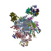
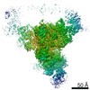
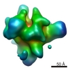
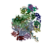
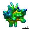
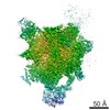
| Title | Post-catalytic P complex spliceosome at 3.3 angstrom resolution from merging data sets | |||||||||
 Map data Map data | None | |||||||||
 Sample Sample |
| |||||||||
| Biological species |  | |||||||||
| Method | single particle reconstruction / cryo EM / Resolution: 3.3 Å | |||||||||
 Authors Authors | Wilkinson ME / Nagai K | |||||||||
| Funding support |  United Kingdom, United Kingdom,  Belgium, 2 items Belgium, 2 items
| |||||||||
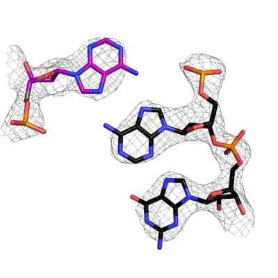
 Citation Citation | Journal: Science / Year: 2017 Title: Postcatalytic spliceosome structure reveals mechanism of 3'-splice site selection. Authors: Max E Wilkinson / Sebastian M Fica / Wojciech P Galej / Christine M Norman / Andrew J Newman / Kiyoshi Nagai / Abstract: Introns are removed from eukaryotic messenger RNA precursors by the spliceosome in two transesterification reactions-branching and exon ligation. The mechanism of 3'-splice site recognition during ...Introns are removed from eukaryotic messenger RNA precursors by the spliceosome in two transesterification reactions-branching and exon ligation. The mechanism of 3'-splice site recognition during exon ligation has remained unclear. Here we present the 3.7-angstrom cryo-electron microscopy structure of the yeast P-complex spliceosome immediately after exon ligation. The 3'-splice site AG dinucleotide is recognized through non-Watson-Crick pairing with the 5' splice site and the branch-point adenosine. After the branching reaction, protein factors work together to remodel the spliceosome and stabilize a conformation competent for 3'-splice site docking, thereby promoting exon ligation. The structure accounts for the strict conservation of the GU and AG dinucleotides at the 5' and 3' ends of introns and provides insight into the catalytic mechanism of exon ligation. | |||||||||
| History |
|
- Structure visualization
| Movie |
 Movie viewer Movie viewer |
|---|---|
| Structure viewer | EM map: SurfViewMolmilJmol/JSmol |
| Supplemental images |
- Downloads & links
-EMDB archive
| Map data | emd_10140.map.gz | 395.7 MB | EMDB map data format | |
|---|---|---|---|---|
| Header (meta data) | emd-10140-v30.xmlemd-10140.xml | 14 KB 14 KB | Display Display | EMDB header |
| FSC (resolution estimation) | emd_10140_fsc.xml | 17 KB | Display | FSC data file |
| Images |  emd_10140.png emd_10140.png | 105 KB | ||
| Archive directory |  http://ftp.pdbj.org/pub/emdb/structures/EMD-10140ftp://ftp.pdbj.org/pub/emdb/structures/EMD-10140 http://ftp.pdbj.org/pub/emdb/structures/EMD-10140ftp://ftp.pdbj.org/pub/emdb/structures/EMD-10140 | HTTPS FTP |
-Related structure data
| Similar structure data | |
|---|---|
| EM raw data | EMPIAR-10298 (Title: Yeast postcatalytic spliceosome, two cryoEM data sets at different magnifications Data size: 6.3 TB Data #1: Unaligned movies of P-complex spliceosome at 105,000x magnification (Dataset 1) [micrographs - multiframe] Data #2: Unaligned movies of P-complex spliceosome at 130,000x magnification (Dataset 2) [micrographs - multiframe]) |


-Links
| EMDB pages | EMDB (EBI/PDBe) / EMDataResource |
|---|
-Map
| File | Download / File: emd_10140.map.gz / Format: CCP4 / Size: 421.9 MB / Type: IMAGE STORED AS FLOATING POINT NUMBER (4 BYTES) | ||||||||||||||||||||||||||||||||||||||||||||||||||||||||||||
|---|---|---|---|---|---|---|---|---|---|---|---|---|---|---|---|---|---|---|---|---|---|---|---|---|---|---|---|---|---|---|---|---|---|---|---|---|---|---|---|---|---|---|---|---|---|---|---|---|---|---|---|---|---|---|---|---|---|---|---|---|---|
| Annotation | None | ||||||||||||||||||||||||||||||||||||||||||||||||||||||||||||
| Projections & slices | Image control
Images are generated by Spider. | ||||||||||||||||||||||||||||||||||||||||||||||||||||||||||||
| Voxel size | X=Y=Z: 1.12 Å | ||||||||||||||||||||||||||||||||||||||||||||||||||||||||||||
| Density |
| ||||||||||||||||||||||||||||||||||||||||||||||||||||||||||||
| Symmetry | Space group: 1 | ||||||||||||||||||||||||||||||||||||||||||||||||||||||||||||
| Details | EMDB XML:
CCP4 map header:
| ||||||||||||||||||||||||||||||||||||||||||||||||||||||||||||
 Z (Sec.)
 Y (Row.)
 X (Col.)


-Supplemental data
- Sample components
-Entire : P complex spliceosome with 3' exon docked
| Entire | Name: P complex spliceosome with 3' exon docked |
|---|---|
| Components |
|
-Supramolecule #1: P complex spliceosome with 3' exon docked
| Supramolecule | Name: P complex spliceosome with 3' exon docked / type: complex / ID: 1 / Parent: 0 |
|---|---|
| Source (natural) | Organism: |
| Molecular weight | Theoretical: 2.2 MDa |
-Experimental details
-Structure determination
| Method | cryo EM |
|---|---|
 Processing Processing | single particle reconstruction |
| Aggregation state | particle |
-Sample preparation
| Concentration | 1.4 mg/mL | ||||||||||||
|---|---|---|---|---|---|---|---|---|---|---|---|---|---|
| Buffer | pH: 7.8 Component:
| ||||||||||||
| Grid | Model: Quantifoil R2/2 / Material: COPPER / Mesh: 400 / Support film - Material: CARBON / Support film - topology: CONTINUOUS / Support film - Film thickness: 7.0 nm / Pretreatment - Type: GLOW DISCHARGE / Pretreatment - Atmosphere: AIR | ||||||||||||
| Vitrification | Cryogen name: ETHANE / Chamber humidity: 100 % / Chamber temperature: 277 K / Instrument: FEI VITROBOT MARK III Details: 3 uL sample was applied to the grid, left for 30s, then blotted for 3s and immediately plunged into liquid ethane. |
- Electron microscopy #1
| Microscopy ID | 1 |
|---|---|
| Microscope | FEI TITAN KRIOS |
| Image recording | Image recording ID: 1 / Film or detector model: GATAN K2 SUMMIT (4k x 4k) / Detector mode: COUNTING / Number grids imaged: 1 / Number real images: 2384 / Average exposure time: 12.0 sec. / Average electron dose: 47.0 e/Å2 |
| Electron beam | Acceleration voltage: 300 kV / Electron source:  FIELD EMISSION GUN FIELD EMISSION GUN |
| Electron optics | Illumination mode: FLOOD BEAM / Imaging mode: BRIGHT FIELD / Cs: 2.7 mm / Nominal defocus max: 3.0 µm / Nominal defocus min: 0.2 µm / Nominal magnification: 105000 |
| Sample stage | Specimen holder model: FEI TITAN KRIOS AUTOGRID HOLDER / Cooling holder cryogen: NITROGEN |
| Experimental equipment |  Model: Titan Krios / Image courtesy: FEI Company |
-Electron microscopy #1~
| Microscopy ID | 1 |
|---|---|
| Microscope | FEI TITAN KRIOS |
| Image recording | #0 - Image recording ID: 2 / #0 - Film or detector model: GATAN K2 SUMMIT (4k x 4k) / #0 - Detector mode: SUPER-RESOLUTION / #0 - Number grids imaged: 1 / #0 - Number real images: 173 / #0 - Average exposure time: 7.0 sec. / #0 - Average electron dose: 45.0 e/Å2 / #1 - Image recording ID: 3 / #1 - Film or detector model: GATAN K2 SUMMIT (4k x 4k) / #1 - Detector mode: COUNTING / #1 - Digitization - Frames/image: 1-35 / #1 - Number grids imaged: 1 / #1 - Number real images: 1441 / #1 - Average exposure time: 7.0 sec. / #1 - Average electron dose: 45.0 e/Å2 |
| Electron beam | Acceleration voltage: 300 kV / Electron source: FIELD EMISSION GUN |
| Electron optics | Illumination mode: FLOOD BEAM / Imaging mode: BRIGHT FIELD / Cs: 2.7 mm / Nominal magnification: 130000 |
| Sample stage | Specimen holder model: FEI TITAN KRIOS AUTOGRID HOLDER / Cooling holder cryogen: NITROGEN |
| Experimental equipment | Model: Titan Krios / Image courtesy: FEI Company |
+Image processing

-Atomic model buiding 1
| Initial model | PDB ID: |
|---|