Movie
Movie Controller


+ Open data

- Basic information
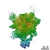
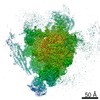
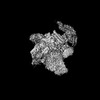
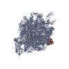
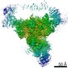
| Entry | Database: EMDB / ID: EMD-3979 | |||||||||
|---|---|---|---|---|---|---|---|---|---|---|
| Title | Post-catalytic P complex spliceosome with 3' splice site docked | |||||||||
 Map data Map data | ||||||||||
 Sample Sample |
| |||||||||
 Keywords Keywords | Spliceosome / P-complex / Exon ligation / SPLICING | |||||||||
| Function / homology |  Function and homology information Function and homology informationU2-type post-spliceosomal complex / mRNA branch site recognition / spliceosomal complex disassembly / U2-type post-mRNA release spliceosomal complex / cellular bud site selection / pre-mRNA 3'-splice site binding / post-mRNA release spliceosomal complex / generation of catalytic spliceosome for first transesterification step / cis assembly of pre-catalytic spliceosome / nuclear mRNA surveillance ...U2-type post-spliceosomal complex / mRNA branch site recognition / spliceosomal complex disassembly / U2-type post-mRNA release spliceosomal complex / cellular bud site selection / pre-mRNA 3'-splice site binding / post-mRNA release spliceosomal complex / generation of catalytic spliceosome for first transesterification step / cis assembly of pre-catalytic spliceosome / nuclear mRNA surveillance / U4/U6 snRNP / 7-methylguanosine cap hypermethylation / spliceosome conformational change to release U4 (or U4atac) and U1 (or U11) / pre-mRNA binding / U2-type catalytic step 1 spliceosome / pICln-Sm protein complex / small nuclear ribonucleoprotein complex / SMN-Sm protein complex / spliceosomal tri-snRNP complex / splicing factor binding / snRNP binding / commitment complex / mRNA cis splicing, via spliceosome / U2-type prespliceosome assembly / U2-type catalytic step 2 spliceosome / U2-type spliceosomal complex / U1 snRNP / U2 snRNP / U4 snRNP / U2-type prespliceosome / poly(U) RNA binding / generation of catalytic spliceosome for second transesterification step / precatalytic spliceosome / mRNA 5'-splice site recognition / mRNA 3'-splice site recognition / spliceosomal complex assembly / Gap-filling DNA repair synthesis and ligation in TC-NER / spliceosomal tri-snRNP complex assembly / Prp19 complex / DNA replication origin binding / U5 snRNP / U5 snRNA binding / Dual incision in TC-NER / pre-mRNA intronic binding / DNA replication initiation / U2 snRNA binding / U6 snRNA binding / protein K63-linked ubiquitination / spliceosomal snRNP assembly / U1 snRNA binding / U4/U6 x U5 tri-snRNP complex / catalytic step 2 spliceosome / positive regulation of cell cycle / nuclear periphery / RNA splicing / positive regulation of RNA splicing / spliceosomal complex / mRNA splicing, via spliceosome / RING-type E3 ubiquitin transferase / metallopeptidase activity / ubiquitin-protein transferase activity / ubiquitin protein ligase activity / RNA helicase activity / RNA helicase / DNA repair / mRNA binding / GTPase activity / chromatin binding / GTP binding / chromatin / ATP hydrolysis activity / mitochondrion / DNA binding / RNA binding / zinc ion binding / ATP binding / metal ion binding / identical protein binding / nucleus / cytosol / cytoplasm Similarity search - Function | |||||||||
| Biological species |  | |||||||||
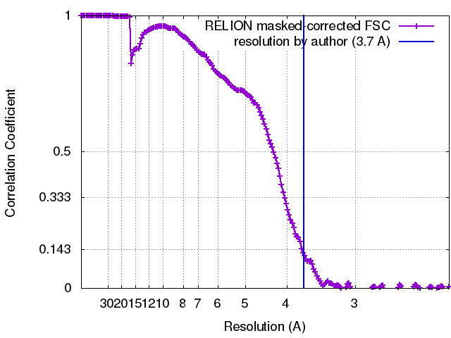
| Method | single particle reconstruction / cryo EM / Resolution: 3.7 Å | |||||||||
 Authors Authors | Wilkinson ME / Fica SM | |||||||||
| Funding support |  United Kingdom, 2 items United Kingdom, 2 items
| |||||||||
 Citation Citation | Journal: Science / Year: 2017 Title: Postcatalytic spliceosome structure reveals mechanism of 3'-splice site selection. Authors: Max E Wilkinson / Sebastian M Fica / Wojciech P Galej / Christine M Norman / Andrew J Newman / Kiyoshi Nagai / Abstract: Introns are removed from eukaryotic messenger RNA precursors by the spliceosome in two transesterification reactions-branching and exon ligation. The mechanism of 3'-splice site recognition during ...Introns are removed from eukaryotic messenger RNA precursors by the spliceosome in two transesterification reactions-branching and exon ligation. The mechanism of 3'-splice site recognition during exon ligation has remained unclear. Here we present the 3.7-angstrom cryo-electron microscopy structure of the yeast P-complex spliceosome immediately after exon ligation. The 3'-splice site AG dinucleotide is recognized through non-Watson-Crick pairing with the 5' splice site and the branch-point adenosine. After the branching reaction, protein factors work together to remodel the spliceosome and stabilize a conformation competent for 3'-splice site docking, thereby promoting exon ligation. The structure accounts for the strict conservation of the GU and AG dinucleotides at the 5' and 3' ends of introns and provides insight into the catalytic mechanism of exon ligation. | |||||||||
| History |
|
- Structure visualization
| Movie |
Movie viewer |
|---|---|
| Structure viewer | EM map: SurfViewMolmilJmol/JSmol |
| Supplemental images |
- Downloads & links
-EMDB archive
| Map data | emd_3979.map.gz | 390.9 MB | EMDB map data format | |
|---|---|---|---|---|
| Header (meta data) | emd-3979-v30.xmlemd-3979.xml | 74.9 KB 74.9 KB | Display Display | EMDB header |
| FSC (resolution estimation) | emd_3979_fsc.xml | 17 KB | Display | FSC data file |
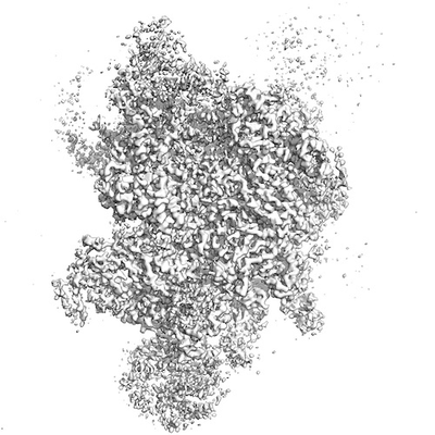
| Images |  emd_3979.png emd_3979.png | 71 KB | ||
| Filedesc metadata | emd-3979.cif.gz | 18.6 KB | ||
| Archive directory |  http://ftp.pdbj.org/pub/emdb/structures/EMD-3979ftp://ftp.pdbj.org/pub/emdb/structures/EMD-3979 http://ftp.pdbj.org/pub/emdb/structures/EMD-3979ftp://ftp.pdbj.org/pub/emdb/structures/EMD-3979 | HTTPS FTP |
-Related structure data
| Related structure data |  6exnMC  3980C M: atomic model generated by this map C: citing same article ( |
|---|---|
| Similar structure data |

-Links
| EMDB pages | EMDB (EBI/PDBe) / EMDataResource |
|---|---|
| Related items in Molecule of the Month |


-Map
| File | Download / File: emd_3979.map.gz / Format: CCP4 / Size: 421.9 MB / Type: IMAGE STORED AS FLOATING POINT NUMBER (4 BYTES) | ||||||||||||||||||||||||||||||||||||||||||||||||||||||||||||
|---|---|---|---|---|---|---|---|---|---|---|---|---|---|---|---|---|---|---|---|---|---|---|---|---|---|---|---|---|---|---|---|---|---|---|---|---|---|---|---|---|---|---|---|---|---|---|---|---|---|---|---|---|---|---|---|---|---|---|---|---|---|
| Projections & slices | Image control
Images are generated by Spider. | ||||||||||||||||||||||||||||||||||||||||||||||||||||||||||||
| Voxel size | X=Y=Z: 1.12 Å | ||||||||||||||||||||||||||||||||||||||||||||||||||||||||||||
| Density |
| ||||||||||||||||||||||||||||||||||||||||||||||||||||||||||||
| Symmetry | Space group: 1 | ||||||||||||||||||||||||||||||||||||||||||||||||||||||||||||
| Details | EMDB XML:
CCP4 map header:
| ||||||||||||||||||||||||||||||||||||||||||||||||||||||||||||
 Z (Sec.)
 Y (Row.)
 X (Col.)


-Supplemental data
- Sample components
+Entire : P complex spliceosome with 3' exon docked
+Supramolecule #1: P complex spliceosome with 3' exon docked
+Supramolecule #2: P complex spliceosome
+Supramolecule #3: 3' exon
+Macromolecule #1: U2 snRNA
+Macromolecule #2: U5 snRNA
+Macromolecule #3: U6 snRNA
+Macromolecule #7: Ligated exons: UBC4 mRNA
+Macromolecule #9: Intron lariat: UBC4 RNA
+Macromolecule #4: Pre-mRNA-splicing factor Prp8
+Macromolecule #5: Pre-mRNA-splicing factor SNU114
+Macromolecule #6: Pre-mRNA-splicing factor CWC16
+Macromolecule #8: Pre-mRNA-splicing factor CWC22
+Macromolecule #10: Pre-mRNA-splicing factor PRP46
+Macromolecule #11: Pre-mRNA-processing protein 45
+Macromolecule #12: Pre-mRNA-splicing factor BUD31
+Macromolecule #13: Pre-mRNA-splicing factor CWC2
+Macromolecule #14: Pre-mRNA-splicing factor SLT11
+Macromolecule #15: Pre-mRNA-splicing factor CEF1
+Macromolecule #16: Pre-mRNA-splicing factor CWC15
+Macromolecule #17: Pre-mRNA-splicing factor CWC21
+Macromolecule #18: Pre-mRNA-splicing factor CLF1
+Macromolecule #19: Pre-mRNA-splicing factor SYF1
+Macromolecule #20: Pre-mRNA-splicing factor ATP-dependent RNA helicase PRP22
+Macromolecule #21: U2 small nuclear ribonucleoprotein A'
+Macromolecule #22: Unassigned structure
+Macromolecule #23: U2 small nuclear ribonucleoprotein B''
+Macromolecule #24: Pre-mRNA-splicing factor Prp18
+Macromolecule #25: Small nuclear ribonucleoprotein-associated protein B
+Macromolecule #26: Pre-mRNA-splicing factor SLU7
+Macromolecule #27: Small nuclear ribonucleoprotein Sm D3
+Macromolecule #28: Small nuclear ribonucleoprotein E
+Macromolecule #29: Small nuclear ribonucleoprotein F
+Macromolecule #30: Small nuclear ribonucleoprotein G
+Macromolecule #31: Small nuclear ribonucleoprotein Sm D1
+Macromolecule #32: Small nuclear ribonucleoprotein Sm D2
+Macromolecule #33: Pre-mRNA-processing factor Prp17
+Macromolecule #34: Pre-mRNA-splicing factor SNT309
+Macromolecule #35: Pre-mRNA-processing factor Prp19
+Macromolecule #36: Pre-mRNA-splicing factor SYF2
+Macromolecule #37: MAGNESIUM ION
+Macromolecule #38: INOSITOL HEXAKISPHOSPHATE

+Macromolecule #39: GUANOSINE-5'-TRIPHOSPHATE

+Macromolecule #40: ZINC ION
-Experimental details
-Structure determination
| Method | cryo EM |
|---|---|
 Processing Processing | single particle reconstruction |
| Aggregation state | particle |
-Sample preparation
| Concentration | 1.4 mg/mL | ||||||||||||
|---|---|---|---|---|---|---|---|---|---|---|---|---|---|
| Buffer | pH: 7.9 Component:
| ||||||||||||
| Grid | Model: Quantifoil R2/2 / Material: COPPER / Mesh: 400 / Support film - Material: CARBON / Support film - topology: CONTINUOUS / Support film - Film thickness: 7 / Pretreatment - Type: GLOW DISCHARGE / Pretreatment - Time: 15 sec. / Pretreatment - Atmosphere: AIR | ||||||||||||
| Vitrification | Cryogen name: ETHANE / Chamber humidity: 100 % / Chamber temperature: 277 K / Instrument: FEI VITROBOT MARK III Details: 3 uL sample was applied to the grid, left for 30s, then blotted for 3s and immediately plunged into liquid ethane.. |
- Electron microscopy
| Microscope | FEI TITAN KRIOS |
|---|---|
| Image recording | Film or detector model: GATAN K2 SUMMIT (4k x 4k) / Detector mode: COUNTING / Digitization - Frames/image: 1-20 / Number grids imaged: 1 / Number real images: 2295 / Average exposure time: 12.0 sec. / Average electron dose: 47.0 e/Å2 |
| Electron beam | Acceleration voltage: 300 kV / Electron source:  FIELD EMISSION GUN FIELD EMISSION GUN |
| Electron optics | C2 aperture diameter: 50.0 µm / Calibrated defocus max: 3.0 µm / Calibrated defocus min: 0.2 µm / Illumination mode: FLOOD BEAM / Imaging mode: BRIGHT FIELD / Cs: 2.7 mm / Nominal magnification: 105000 |
| Sample stage | Specimen holder model: FEI TITAN KRIOS AUTOGRID HOLDER / Cooling holder cryogen: NITROGEN |
| Experimental equipment |  Model: Titan Krios / Image courtesy: FEI Company |