Movie
Movie Controller


+ Open data

- Basic information
| Entry | Database: EMDB / ID: EMD-10113 | |||||||||
|---|---|---|---|---|---|---|---|---|---|---|
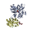
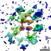
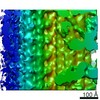
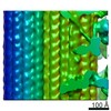
| Title | Basketweave Z-band of sonic muscle in midshipman fish | |||||||||
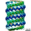
 Map data Map data | Subtomogram average of a tomogram of basketweave Z-band found in sonic muscle in the swimbladder of male Type 1 midshipman fish. | |||||||||
 Sample Sample |
| |||||||||
| Biological species |  Porichthys notatus (plainfin midshipman) Porichthys notatus (plainfin midshipman) | |||||||||
| Method | subtomogram averaging / cryo EM / negative staining / Resolution: 39.0 Å | |||||||||
 Authors Authors | Luther PK / Heumann J / Burgoyne T / Morris EP | |||||||||
| Funding support |  United Kingdom, 1 items United Kingdom, 1 items
| |||||||||
 Citation Citation | Journal: Proc Natl Acad Sci U S A / Year: 2019 Title: Three-dimensional structure of the basketweave Z-band in midshipman fish sonic muscle. Authors: Thomas Burgoyne / John M Heumann / Edward P Morris / Carlo Knupp / Jun Liu / Michael K Reedy / Kenneth A Taylor / Kuan Wang / Pradeep K Luther /   Abstract: Striated muscle enables movement in all animals by the contraction of myriads of sarcomeres joined end to end by the Z-bands. The contraction is due to tension generated in each sarcomere between ...Striated muscle enables movement in all animals by the contraction of myriads of sarcomeres joined end to end by the Z-bands. The contraction is due to tension generated in each sarcomere between overlapping arrays of actin and myosin filaments. At the Z-band, actin filaments from adjoining sarcomeres overlap and are cross-linked in a regular pattern mainly by the protein α-actinin. The Z-band is dynamic, reflected by the 2 regular patterns seen in transverse section electron micrographs; the so-called small-square and basketweave forms. Although these forms are attributed, respectively, to relaxed and actively contracting muscles, the basketweave form occurs in certain relaxed muscles as in the muscle studied here. We used electron tomography and subtomogram averaging to derive the 3D structure of the Z-band in the swimbladder sonic muscle of type I male plainfin midshipman fish (, into which we docked the crystallographic structures of actin and α-actinin. The α-actinin links run diagonally between connected pairs of antiparallel actin filaments and are oriented at an angle of about 25° away from the actin filament axes. The slightly curved and flattened structure of the α-actinin rod has a distinct fit into the map. The Z-band model provides a detailed understanding of the role of α-actinin in transmitting tension between actin filaments in adjoining sarcomeres. | |||||||||
| History |
|
- Structure visualization
| Movie |
 Movie viewer Movie viewer |
|---|---|
| Structure viewer | EM map: SurfViewMolmilJmol/JSmol |
| Supplemental images |
- Downloads & links
-EMDB archive
| Map data | emd_10113.map.gz | 3.9 MB | EMDB map data format | |
|---|---|---|---|---|
| Header (meta data) | emd-10113-v30.xmlemd-10113.xml | 14.4 KB 14.4 KB | Display Display | EMDB header |
| Images |  emd_10113.png emd_10113.png | 59.3 KB | ||
| Archive directory |  http://ftp.pdbj.org/pub/emdb/structures/EMD-10113ftp://ftp.pdbj.org/pub/emdb/structures/EMD-10113 http://ftp.pdbj.org/pub/emdb/structures/EMD-10113ftp://ftp.pdbj.org/pub/emdb/structures/EMD-10113 | HTTPS FTP |
-Related structure data
| Similar structure data |
|---|

-Links
| EMDB pages | EMDB (EBI/PDBe) / EMDataResource |
|---|
-Map
| File | Download / File: emd_10113.map.gz / Format: CCP4 / Size: 4.9 MB / Type: IMAGE STORED AS FLOATING POINT NUMBER (4 BYTES) | ||||||||||||||||||||||||||||||||||||||||||||||||||||||||||||||||||||
|---|---|---|---|---|---|---|---|---|---|---|---|---|---|---|---|---|---|---|---|---|---|---|---|---|---|---|---|---|---|---|---|---|---|---|---|---|---|---|---|---|---|---|---|---|---|---|---|---|---|---|---|---|---|---|---|---|---|---|---|---|---|---|---|---|---|---|---|---|---|
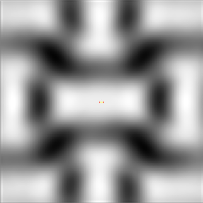
| Annotation | Subtomogram average of a tomogram of basketweave Z-band found in sonic muscle in the swimbladder of male Type 1 midshipman fish. | ||||||||||||||||||||||||||||||||||||||||||||||||||||||||||||||||||||
| Projections & slices | Image control
Images are generated by Spider. generated in cubic-lattice coordinate | ||||||||||||||||||||||||||||||||||||||||||||||||||||||||||||||||||||
| Voxel size | X=Y=Z: 6.42 Å | ||||||||||||||||||||||||||||||||||||||||||||||||||||||||||||||||||||
| Density |
| ||||||||||||||||||||||||||||||||||||||||||||||||||||||||||||||||||||
| Symmetry | Space group: 1 | ||||||||||||||||||||||||||||||||||||||||||||||||||||||||||||||||||||
| Details | EMDB XML:
CCP4 map header:
| ||||||||||||||||||||||||||||||||||||||||||||||||||||||||||||||||||||
 Z (Sec.)
 Y (Row.)
 X (Col.)


-Supplemental data
- Sample components
-Entire : Z-band of vertebrate striated muscle in basketweave form
| Entire | Name: Z-band of vertebrate striated muscle in basketweave form |
|---|---|
| Components |
|
-Supramolecule #1: Z-band of vertebrate striated muscle in basketweave form
| Supramolecule | Name: Z-band of vertebrate striated muscle in basketweave form type: organelle_or_cellular_component / ID: 1 / Parent: 0 Details: Z-band studied in sonic muscle in the swimbladder of male Type 1 midshipman fish. |
|---|---|
| Source (natural) | Organism: Porichthys notatus (plainfin midshipman) / Organ: Swimbladder muscle / Tissue: Muscle / Organelle: Z-band / Location in cell: Myofibrillar |
-Experimental details
-Structure determination
| Method | negative staining, cryo EM |
|---|---|
 Processing Processing | subtomogram averaging |
| Aggregation state | tissue |
-Sample preparation
| Buffer | pH: 7 Component:
Details: This is the relaxing solution | ||||||||||||||||||||||||
|---|---|---|---|---|---|---|---|---|---|---|---|---|---|---|---|---|---|---|---|---|---|---|---|---|---|
| Staining | Type: POSITIVE / Material: Uranyl acetate and lead citrate Details: Conventionally stained with 2% uranyl acetate and Reynolds lead citrate | ||||||||||||||||||||||||
| Sugar embedding | Material: Araldite Details: Sample was freeze-substituted in acetone and then embedded in araldiate | ||||||||||||||||||||||||
| Grid | Material: COPPER / Mesh: 700 | ||||||||||||||||||||||||
| Vitrification | Cryogen name: ETHANE / Instrument: REICHERT-JUNG PLUNGER Details: Sample was cryo-protected with 30% sucrose (added to the relaxing solution). | ||||||||||||||||||||||||
| Details | Fibers of swimbladder muscle (sonic muscle) of midshipman fish were rapidly frozen, freeze-substituted, embedded in resin, ~100 nm thin transverse sections cut, coated with 10 nm gold fiducials and stained with uranyl acetate and lead citrate. |
- Electron microscopy
| Microscope | FEI/PHILIPS CM200FEG |
|---|---|
| Image recording | Film or detector model: TVIPS TEMCAM-F415 (4k x 4k) / Digitization - Dimensions - Width: 4096 pixel / Digitization - Dimensions - Height: 4096 pixel / Number grids imaged: 1 / Average exposure time: 1.0 sec. / Average electron dose: 5.0 e/Å2 |
| Electron beam | Acceleration voltage: 200 kV / Electron source:  FIELD EMISSION GUN FIELD EMISSION GUN |
| Electron optics | Illumination mode: FLOOD BEAM / Imaging mode: BRIGHT FIELD / Nominal defocus min: 0.005 µm / Nominal magnification: 27000 |
| Sample stage | Specimen holder model: OTHER / Cooling holder cryogen: NITROGEN |