 Movie
Movie Controller

[English] 日本語
 Yorodumi
- EMDB-0629: The axonemal doublet microtubule focusing on the inner junction r... -
+ Open data

- Basic information
| Entry | Database: EMDB / ID: EMD-0629 | ||||||||||||
|---|---|---|---|---|---|---|---|---|---|---|---|---|---|
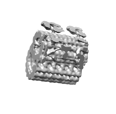
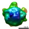
| Title | The axonemal doublet microtubule focusing on the inner junction region extracted from the cryo-electron tomography and subtomographic average of isolated Chlamydomonas pacrg mutant cilia | ||||||||||||
 Map data Map data | The axonemal doublet microtubule focusing on the inner junction region extracted from the cryo-electron tomography and subtomographic average of isolated Chlamydomonas pacrg mutant cilia | ||||||||||||
 Sample Sample |
| ||||||||||||
| Biological species |   Chlamydomonas reinhardtii (plant) Chlamydomonas reinhardtii (plant) | ||||||||||||
| Method | subtomogram averaging / cryo EM / Resolution: 27.0 Å | ||||||||||||
 Authors Authors | Lin J / Fu G / Nicastro D / Dymek EE / Porter M / Smith EF | ||||||||||||
| Funding support |  United States, 3 items United States, 3 items
| ||||||||||||
 Citation Citation | Journal: Mol Biol Cell / Year: 2019 Title: PACRG and FAP20 form the inner junction of axonemal doublet microtubules and regulate ciliary motility. Authors: Erin E Dymek / Jianfeng Lin / Gang Fu / Mary E Porter / Daniela Nicastro / Elizabeth F Smith / Abstract: We previously demonstrated that PACRG plays a role in regulating dynein-driven microtubule sliding in motile cilia. To expand our understanding of the role of PACRG in ciliary assembly and motility, ...We previously demonstrated that PACRG plays a role in regulating dynein-driven microtubule sliding in motile cilia. To expand our understanding of the role of PACRG in ciliary assembly and motility, we used a combination of functional and structural studies, including newly identified mutants. Using cryo-electron tomography we show that PACRG and FAP20 form the inner junction between the A- and B-tubule along the length of all nine ciliary doublet microtubules. The lack of PACRG and FAP20 also results in reduced assembly of inner-arm dynein IDA and the beak-MIP structures. In addition, our functional studies reveal that loss of PACRG and/or FAP20 causes severe cell motility defects and reduced in vitro microtubule sliding velocities. Interestingly, the addition of exogenous PACRG and/or FAP20 protein to isolated mutant axonemes restores microtubule sliding velocities, but not ciliary beating. Taken together, these studies show that PACRG and FAP20 comprise the inner junction bridge that serves as a hub for both directly modulating dynein-driven microtubule sliding, as well as for the assembly of additional ciliary components that play essential roles in generating coordinated ciliary beating. | ||||||||||||
| History |
|
- Structure visualization
| Movie |
 Movie viewer Movie viewer |
|---|---|
| Structure viewer | EM map: SurfViewMolmilJmol/JSmol |
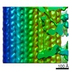
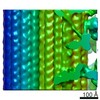
| Supplemental images |
- Downloads & links
-EMDB archive
| Map data | emd_0629.map.gz | 1.5 MB | EMDB map data format | |
|---|---|---|---|---|
| Header (meta data) | emd-0629-v30.xmlemd-0629.xml | 11.8 KB 11.8 KB | Display Display | EMDB header |
| Images |  emd_0629.png emd_0629.png | 58.5 KB | ||
| Archive directory |  http://ftp.pdbj.org/pub/emdb/structures/EMD-0629ftp://ftp.pdbj.org/pub/emdb/structures/EMD-0629 http://ftp.pdbj.org/pub/emdb/structures/EMD-0629ftp://ftp.pdbj.org/pub/emdb/structures/EMD-0629 | HTTPS FTP |
-Related structure data
-Links
| EMDB pages | EMDB (EBI/PDBe) / EMDataResource |
|---|
-Map
| File | Download / File: emd_0629.map.gz / Format: CCP4 / Size: 2 MB / Type: IMAGE STORED AS FLOATING POINT NUMBER (4 BYTES) | ||||||||||||||||||||||||||||||||||||||||||||||||||||||||||||
|---|---|---|---|---|---|---|---|---|---|---|---|---|---|---|---|---|---|---|---|---|---|---|---|---|---|---|---|---|---|---|---|---|---|---|---|---|---|---|---|---|---|---|---|---|---|---|---|---|---|---|---|---|---|---|---|---|---|---|---|---|---|
| Annotation | The axonemal doublet microtubule focusing on the inner junction region extracted from the cryo-electron tomography and subtomographic average of isolated Chlamydomonas pacrg mutant cilia | ||||||||||||||||||||||||||||||||||||||||||||||||||||||||||||
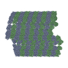
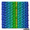
| Projections & slices | Image control
Images are generated by Spider. generated in cubic-lattice coordinate | ||||||||||||||||||||||||||||||||||||||||||||||||||||||||||||
| Voxel size | X=Y=Z: 5.523 Å | ||||||||||||||||||||||||||||||||||||||||||||||||||||||||||||
| Density |
| ||||||||||||||||||||||||||||||||||||||||||||||||||||||||||||
| Symmetry | Space group: 1 | ||||||||||||||||||||||||||||||||||||||||||||||||||||||||||||
| Details | EMDB XML:
CCP4 map header:
| ||||||||||||||||||||||||||||||||||||||||||||||||||||||||||||
 Z (Sec.)
 Y (Row.)
 X (Col.)


-Supplemental data
- Sample components
-Entire : The inner junction region of axonemal doublet microtubule average...
| Entire | Name: The inner junction region of axonemal doublet microtubule averaged from Chlamydomonas pacrg mutant cilia |
|---|---|
| Components |
|
-Supramolecule #1: The inner junction region of axonemal doublet microtubule average...
| Supramolecule | Name: The inner junction region of axonemal doublet microtubule averaged from Chlamydomonas pacrg mutant cilia type: organelle_or_cellular_component / ID: 1 / Parent: 0 |
|---|---|
| Source (natural) | Organism: Chlamydomonas reinhardtii (plant) / Organelle: cilia |
-Experimental details
-Structure determination
| Method | cryo EM |
|---|---|
 Processing Processing | subtomogram averaging |
| Aggregation state | cell |
-Sample preparation
| Buffer | pH: 7.4 |
|---|---|
| Grid | Support film - Material: CARBON / Support film - topology: HOLEY / Details: unspecified |
| Vitrification | Cryogen name: ETHANE / Chamber temperature: 298 K / Instrument: HOMEMADE PLUNGER Details: back-side blotting for 1.5-2.5 seconds before plunging. |
| Details | Freshly isolated and demembranated cilia from Chlamydomonas pacrg mutant cells |
- Electron microscopy
| Microscope | FEI TITAN KRIOS |
|---|---|
| Specialist optics | Phase plate: VOLTA PHASE PLATE / Energy filter - Name: GIF Bioquantum / Energy filter - Slit width: 20 eV |
| Image recording | Film or detector model: GATAN K2 SUMMIT (4k x 4k) / Detector mode: COUNTING / Digitization - Dimensions - Width: 3710 pixel / Digitization - Dimensions - Height: 3838 pixel / Digitization - Frames/image: 1-15 / Average exposure time: 6.0 sec. / Average electron dose: 1.57 e/Å2 |
| Electron beam | Acceleration voltage: 300 kV / Electron source:  FIELD EMISSION GUN FIELD EMISSION GUN |
| Electron optics | C2 aperture diameter: 100.0 µm / Calibrated defocus max: 0.5 µm / Calibrated defocus min: 0.2 µm / Calibrated magnification: 26000 / Illumination mode: FLOOD BEAM / Imaging mode: BRIGHT FIELD |
| Sample stage | Specimen holder model: FEI TITAN KRIOS AUTOGRID HOLDER / Cooling holder cryogen: NITROGEN |
| Experimental equipment |  Model: Titan Krios / Image courtesy: FEI Company |
-Image processing
| Final reconstruction | Applied symmetry - Point group: C1 (asymmetric) / Resolution.type: BY AUTHOR / Resolution: 27.0 Å / Resolution method: FSC 0.5 CUT-OFF / Software - Name: IMOD / Number subtomograms used: 2333 |
|---|---|
| Extraction | Number tomograms: 19 / Number images used: 2333 / Software - Name: MATLAB |
| Final angle assignment | Type: OTHER / Software - Name: IMOD |
