 Movie
Movie Controller


+ Open data

- Basic information
| Entry | Database: PDB / ID: 3jao | ||||||
|---|---|---|---|---|---|---|---|
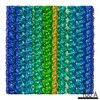
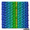
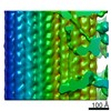
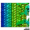
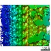
| Title | Ciliary microtubule doublet | ||||||
 Components Components |
| ||||||
 Keywords Keywords | STRUCTURAL PROTEIN / tubulin / microtubule doublet / cilia | ||||||
| Function / homology |  Function and homology information Function and homology informationmicrotubule-based process / structural constituent of cytoskeleton / microtubule / hydrolase activity / GTPase activity / GTP binding / metal ion binding Similarity search - Function | ||||||
| Biological species |   Tetrahymena thermophila (eukaryote) Tetrahymena thermophila (eukaryote) | ||||||
| Method | ELECTRON MICROSCOPY / single particle reconstruction / cryo EM / Resolution: 23 Å | ||||||
 Authors Authors | Maheshwari, A. / Obbineni, J.M. / Bui, K.H. / Shibata, K. / Toyoshima, Y.Y. / Ishikawa, T. | ||||||
 Citation Citation | Journal: Structure / Year: 2015 Title: α- and β-Tubulin Lattice of the Axonemal Microtubule Doublet and Binding Proteins Revealed by Single Particle Cryo-Electron Microscopy and Tomography. Authors: Aditi Maheshwari / Jagan Mohan Obbineni / Khanh Huy Bui / Keitaro Shibata / Yoko Y Toyoshima / Takashi Ishikawa /   Abstract: Microtubule doublet (MTD) is the main skeleton of cilia/flagella. Many proteins, such as dyneins and radial spokes, bind to MTD, and generate or regulate force. While the structure of the ...Microtubule doublet (MTD) is the main skeleton of cilia/flagella. Many proteins, such as dyneins and radial spokes, bind to MTD, and generate or regulate force. While the structure of the reconstituted microtubule has been solved at atomic resolution, nature of the axonemal MTD is still unclear. There are a few hypotheses of the lattice arrangement of its α- and β-tubulins, but it has not been described how dyneins and radial spokes bind to MTD. In this study, we analyzed the three-dimensional structure of Tetrahymena MTD at ∼19 Å resolution by single particle cryo-electron microscopy. To identify α- and β-tubulins, we combined image analysis of MTD with specific kinesin decoration. This work reveals that α- and β-tubulins form a B-lattice arrangement in the entire MTD with a seam at the outer junction. We revealed the unique way in which inner arm dyneins, radial spokes, and proteins inside MTD bind and bridge protofilaments. | ||||||
| History |
|
- Structure visualization
| Movie |
Movie viewer |
|---|---|
| Structure viewer | Molecule: MolmilJmol/JSmol |
- Downloads & links
-Download
| PDBx/mmCIF format | 3jao.cif.gz | 170.3 KB | Display | PDBx/mmCIF format |
|---|---|---|---|---|
| PDB format | pdb3jao.ent.gz | 128.4 KB | Display | PDB format |
| PDBx/mmJSON format | 3jao.json.gz | Tree view | PDBx/mmJSON format | |
| Others |  Other downloads Other downloads |
-Validation report
| Arichive directory | https://data.pdbj.org/pub/pdb/validation_reports/ja/3jaoftp://data.pdbj.org/pub/pdb/validation_reports/ja/3jao | HTTPS FTP |
|---|
-Related structure data
| Related structure data |  6312MC M: map data used to model this data C: citing same article ( |
|---|---|
| Similar structure data |
-Links
 PDBj
PDBj


- Assembly
| Deposited unit | 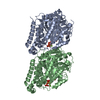
|
|---|---|
| 1 | x 115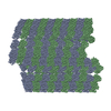
|
| 2 |
|
| 3 | 
|
-Components
-Protein , 2 types, 2 molecules AB
| #1: Protein | Mass: 50107.238 Da / Num. of mol.: 1 / Fragment: SEE REMARK 999 / Source method: isolated from a natural source / Source: (natural) Tetrahymena thermophila (eukaryote) / Strain: SB210 / References: UniProt: A0A0M3KKT1*PLUS |
|---|---|
| #2: Protein | Mass: 49907.770 Da / Num. of mol.: 1 / Fragment: SEE REMARK 999 / Source method: isolated from a natural source / Source: (natural) Tetrahymena thermophila (eukaryote) / Strain: SB210 / References: UniProt: A0A0M3KKT2*PLUS |
-Non-polymers , 4 types, 12 molecules 


| #3: Chemical | ChemComp-GTP /  Mass: 523.180 Da / Num. of mol.: 1 / Source method: obtained synthetically / Formula: C10H16N5O14P3 / Comment: GTP, energy-carrying molecule*YM Mass: 523.180 Da / Num. of mol.: 1 / Source method: obtained synthetically / Formula: C10H16N5O14P3 / Comment: GTP, energy-carrying molecule*YM | ||||
|---|---|---|---|---|---|
| #4: Chemical |  Mass: 24.305 Da / Num. of mol.: 2 / Source method: obtained synthetically / Formula: Mg Mass: 24.305 Da / Num. of mol.: 2 / Source method: obtained synthetically / Formula: Mg#5: Chemical | ChemComp-G2P / |  Mass: 521.208 Da / Num. of mol.: 1 / Source method: obtained synthetically / Formula: C11H18N5O13P3 / Comment: GMP-CPP, energy-carrying molecule analogue*YM Mass: 521.208 Da / Num. of mol.: 1 / Source method: obtained synthetically / Formula: C11H18N5O13P3 / Comment: GMP-CPP, energy-carrying molecule analogue*YM#6: Water | ChemComp-HOH / | Mass: 18.015 Da / Num. of mol.: 8 / Source method: isolated from a natural source / Formula: H2O |
-Details
| Sequence details | MICROTUBULES FROM TETRAHYMENA THERMOPHILA WERE IMAGED, BUT PROTEIN SEQUENCES FROM SUS SCROFA (UNP ...MICROTUBUL |
|---|
-Experimental details
-Experiment
| Experiment | Method: ELECTRON MICROSCOPY |
|---|---|
| EM experiment | Aggregation state: FILAMENT / 3D reconstruction method: single particle reconstruction |
- Sample preparation
| Component | Name: microtubule doublet extracted from Tetrahymena cilia / Type: COMPLEX |
|---|---|
| Buffer solution | Name: HMDEK buffer / pH: 7.4 Details: 30 mM HEPES, pH 7.4, 5 mM MgSO4, 1 mM DTT, 0.5 mM EDTA, 25 mM KCl, 4 mM CaCl2 |
| Specimen | Conc.: 3 mg/ml / Embedding applied: NO / Shadowing applied: NO / Staining applied: NO / Vitrification applied: YES |
| Specimen support | Details: 300 mesh copper grid with holey carbon film, glow discharged |
| Vitrification | Instrument: FEI VITROBOT MARK II / Cryogen name: ETHANE / Temp: 90 K / Humidity: 90 % Details: Blot for 3 seconds in humid air atmosphere before plunging into liquid ethane (FEI VITROBOT MARK II). Method: Blot for 3 seconds before plunging |
- Electron microscopy imaging
| Experimental equipment |  Model: Tecnai F20 / Image courtesy: FEI Company |
|---|---|
| Microscopy | Model: FEI TECNAI F20 / Date: Oct 25, 2011 |
| Electron gun | Electron source:  FIELD EMISSION GUN / Accelerating voltage: 200 kV / Illumination mode: FLOOD BEAM FIELD EMISSION GUN / Accelerating voltage: 200 kV / Illumination mode: FLOOD BEAM |
| Electron lens | Mode: BRIGHT FIELD / Nominal magnification: 67000 X / Calibrated magnification: 67000 X / Nominal defocus max: 4000 nm / Nominal defocus min: 1000 nm / Cs: 1.5 mm |
| Specimen holder | Specimen holder model: GATAN LIQUID NITROGEN / Temperature: 90 K / Temperature (max): 100 K / Temperature (min): 80 K |
| Image recording | Electron dose: 30 e/Å2 / Film or detector model: GENERIC CCD / Details: Gatan Ultrascan 4000 |
| EM imaging optics | Energyfilter name: Gatan GIF Tridem Energy Imaging Filter / Energyfilter upper: 25 eV / Energyfilter lower: 0 eV |
| Radiation | Protocol: SINGLE WAVELENGTH / Monochromatic (M) / Laue (L): M / Scattering type: x-ray |
| Radiation wavelength | Relative weight: 1 |
- Processing
| EM software |
| ||||||||||||
|---|---|---|---|---|---|---|---|---|---|---|---|---|---|
| CTF correction | Details: each particle | ||||||||||||
| 3D reconstruction | Method: R-weighted back projection / Resolution: 23 Å / Resolution method: FSC 0.143 CUT-OFF / Num. of particles: 10700 / Nominal pixel size: 4.4776 Å / Actual pixel size: 4.4776 Å / Details: gold standard / Refinement type: HALF-MAPS REFINED INDEPENDENTLY / Symmetry type: HELICAL | ||||||||||||
| Atomic model building | Protocol: RIGID BODY FIT / Space: REAL / Target criteria: Cross-correlation coefficient Details: METHOD--kinesin binding, gradient density map analysis, and cross-correlation coefficient. The fitting for protofilament A10 as represented by BIOMT records 51-55 is ambiguous as it has been ...Details: METHOD--kinesin binding, gradient density map analysis, and cross-correlation coefficient. The fitting for protofilament A10 as represented by BIOMT records 51-55 is ambiguous as it has been assigned using only the cross-correlation coefficient. REFINEMENT PROTOCOL--RIGID BODY DETAILS--rigid body fitting of individual dimers | ||||||||||||
| Atomic model building | PDB-ID: 3J6E Accession code: 3J6E / Source name: PDB / Type: experimental model | ||||||||||||
| Refinement step | Cycle: LAST
|

