Movie
Movie Controller

[English] 日本語
 Yorodumi
- EMDB-1152: Electron cryotomography of the E. coli pyruvate and 2-oxoglutarat... -
+ Open data

- Basic information
| Entry | Database: EMDB / ID: EMD-1152 | |||||||||
|---|---|---|---|---|---|---|---|---|---|---|
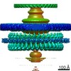
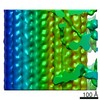
| Title | Electron cryotomography of the E. coli pyruvate and 2-oxoglutarate dehydrogenase complexes. | |||||||||
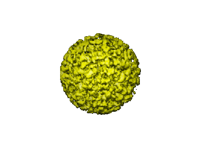
 Map data Map data | Volume of an extracted E. coli 2-oxoglutarate multienzyme complex particle from a dual-tilt tomogram. | |||||||||
 Sample Sample |
| |||||||||
| Function / homology | Dihydrolipoamide succinyltransferase / 2-oxoglutarate dehydrogenase E1 component / Dihydrolipoamide dehydrogenase Function and homology information Function and homology information | |||||||||
| Biological species |  | |||||||||
| Method | subtomogram averaging / cryo EM / Resolution: 55.0 Å | |||||||||
 Authors Authors | Murphy GE / Jensen GJ | |||||||||
 Citation Citation | Journal: Structure / Year: 2005 Title: Electron cryotomography of the E. coli pyruvate and 2-oxoglutarate dehydrogenase complexes. Authors: Gavin E Murphy / Grant J Jensen /  Abstract: The E. coli pyruvate and 2-oxoglutarate dehydrogenases are two closely related, large complexes that exemplify a growing number of multiprotein "machines" whose domains have been studied extensively ...The E. coli pyruvate and 2-oxoglutarate dehydrogenases are two closely related, large complexes that exemplify a growing number of multiprotein "machines" whose domains have been studied extensively and modeled in atomic detail, but whose quaternary structures have remained unclear for lack of an effective imaging technology. Here, electron cryotomography was used to show that the E1 and E3 subunits of these complexes are flexibly tethered approximately 11 nm away from the E2 core. This result demonstrates unambiguously that electron cryotomography can reveal the relative positions of features as small as 80 kDa in individual complexes, elucidating quaternary structure and conformational flexibility. | |||||||||
| History |
|
- Structure visualization
| Movie |
Movie viewer |
|---|---|
| Structure viewer | EM map: SurfViewMolmilJmol/JSmol |
| Supplemental images |
- Downloads & links
-EMDB archive
| Map data | emd_1152.map.gz | 227 KB | EMDB map data format | |
|---|---|---|---|---|
| Header (meta data) | emd-1152-v30.xmlemd-1152.xml | 12.9 KB 12.9 KB | Display Display | EMDB header |
| Images |  1152.gif 1152.gif | 23.6 KB | ||
| Archive directory |  http://ftp.pdbj.org/pub/emdb/structures/EMD-1152ftp://ftp.pdbj.org/pub/emdb/structures/EMD-1152 http://ftp.pdbj.org/pub/emdb/structures/EMD-1152ftp://ftp.pdbj.org/pub/emdb/structures/EMD-1152 | HTTPS FTP |
-Related structure data

-Links
| EMDB pages | EMDB (EBI/PDBe) / EMDataResource |
|---|
-Map
| File | Download / File: emd_1152.map.gz / Format: CCP4 / Size: 977.5 KB / Type: IMAGE STORED AS SIGNED INTEGER (2 BYTES) | ||||||||||||||||||||||||||||||||||||||||||||||||||||||||||||||||||||
|---|---|---|---|---|---|---|---|---|---|---|---|---|---|---|---|---|---|---|---|---|---|---|---|---|---|---|---|---|---|---|---|---|---|---|---|---|---|---|---|---|---|---|---|---|---|---|---|---|---|---|---|---|---|---|---|---|---|---|---|---|---|---|---|---|---|---|---|---|---|
| Annotation | Volume of an extracted E. coli 2-oxoglutarate multienzyme complex particle from a dual-tilt tomogram. | ||||||||||||||||||||||||||||||||||||||||||||||||||||||||||||||||||||
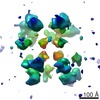
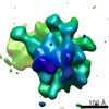
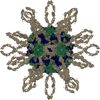
| Projections & slices | Image control
Images are generated by Spider. | ||||||||||||||||||||||||||||||||||||||||||||||||||||||||||||||||||||
| Voxel size | X=Y=Z: 8.2 Å | ||||||||||||||||||||||||||||||||||||||||||||||||||||||||||||||||||||
| Density |
| ||||||||||||||||||||||||||||||||||||||||||||||||||||||||||||||||||||
| Symmetry | Space group: 1 | ||||||||||||||||||||||||||||||||||||||||||||||||||||||||||||||||||||
| Details | EMDB XML:
CCP4 map header:
| ||||||||||||||||||||||||||||||||||||||||||||||||||||||||||||||||||||
 Z (Sec.)
 Y (Row.)
 X (Col.)


-Supplemental data
- Sample components
-Entire : Unengineered E. coli 2-oxoglutarate dehydrogenase
| Entire | Name: Unengineered E. coli 2-oxoglutarate dehydrogenase |
|---|---|
| Components |
|
-Supramolecule #1000: Unengineered E. coli 2-oxoglutarate dehydrogenase
| Supramolecule | Name: Unengineered E. coli 2-oxoglutarate dehydrogenase / type: sample / ID: 1000 Details: The sample was thawed from storage at -80 degrees Celcius before being loaded onto the grid. Oligomeric state: Up to 24 E1o 2-oxoglutarate dehydrogenase and E3 dihydrolipoamide dehydrogenase dimers together bind to the 24 dihydrolipoamide succinate transferase E2o octahedral core. Number unique components: 3 |
|---|---|
| Molecular weight | Theoretical: 5.2 MDa |
-Macromolecule #1: E2o octahedral core
| Macromolecule | Name: E2o octahedral core / type: protein_or_peptide / ID: 1 / Name.synonym: dihydrolipoamide succinate transferase / Number of copies: 24 / Oligomeric state: 24mer / Recombinant expression: Yes |
|---|---|
| Source (natural) | Organism: |
| Molecular weight | Experimental: 44 KDa |
| Recombinant expression | Organism: |
| Sequence | InterPro: Dihydrolipoamide succinyltransferase |
-Macromolecule #2: E1o
| Macromolecule | Name: E1o / type: protein_or_peptide / ID: 2 / Name.synonym: 2-oxoglutarate dehydrogenase / Oligomeric state: Dimer / Recombinant expression: Yes |
|---|---|
| Source (natural) | Organism: |
| Molecular weight | Experimental: 200 KDa |
| Recombinant expression | Organism: |
| Sequence | InterPro: 2-oxoglutarate dehydrogenase E1 component |
-Macromolecule #3: E3
| Macromolecule | Name: E3 / type: protein_or_peptide / ID: 3 / Name.synonym: dihydrolipoamide dehydrogenase / Oligomeric state: Dimer / Recombinant expression: Yes |
|---|---|
| Source (natural) | Organism: |
| Molecular weight | Experimental: 100 KDa |
| Recombinant expression | Organism: |
| Sequence | InterPro: Dihydrolipoamide dehydrogenase |
-Experimental details
-Structure determination
| Method | cryo EM |
|---|---|
 Processing Processing | subtomogram averaging |
-Sample preparation
| Concentration | 2 mg/mL |
|---|---|
| Buffer | pH: 7 / Details: 20 mM Potassium Phosphate |
| Grid | Details: R 1.5/1.3 Quantifoil |
| Vitrification | Cryogen name: ETHANE / Chamber humidity: 100 % / Instrument: OTHER Details: Vitrification instrument: Vitrobot. 4 ul of sample placed on grid at 22 C in air before plunging. Method: Blot for 3.5 seconds with an offset of -3 before plunging. |
- Electron microscopy
| Microscope | FEI TECNAI F30 |
|---|---|
| Temperature | Min: 82 K / Max: 82 K / Average: 82 K |
| Alignment procedure | Legacy - Electron beam tilt params: 0 |
| Specialist optics | Energy filter - Name: GIF 3000 / Energy filter - Lower energy threshold: 0.0 eV / Energy filter - Upper energy threshold: 20.0 eV |
| Date | Sep 7, 2004 |
| Image recording | Category: CCD / Film or detector model: GATAN ULTRASCAN 1000 (2k x 2k) / Average electron dose: 120 e/Å2 |
| Electron beam | Acceleration voltage: 300 kV / Electron source:  FIELD EMISSION GUN FIELD EMISSION GUN |
| Electron optics | Calibrated magnification: 36600 / Illumination mode: FLOOD BEAM / Imaging mode: BRIGHT FIELD / Cs: 2 mm / Nominal defocus max: 10.0 µm / Nominal defocus min: 10.0 µm / Nominal magnification: 27500 |
| Sample stage | Specimen holder: FEI Polara / Specimen holder model: GATAN HELIUM / Tilt series - Axis1 - Min angle: -63 ° / Tilt series - Axis1 - Max angle: 63 ° |
| Experimental equipment |  Model: Tecnai F30 / Image courtesy: FEI Company |
-Image processing
| Details | Dual-axis tilt series, with 44 sections on one axis, and 44 on the other. Average number of tilts used in the 3D reconstructions: 88. Average tomographic tilt angle increment: 3. |
|---|---|
| Final reconstruction | Algorithm: OTHER / Resolution.type: BY AUTHOR / Resolution: 55.0 Å / Resolution method: FSC 0.5 CUT-OFF / Software - Name: IMOD Details: The individual, orthogonal tomograms were filtered at their first CTF zero and then merged. |
-Atomic model buiding 1
| Initial model | PDB ID: |
|---|---|
| Details | The octameric E2o core (1E2O) was fit into the core density using bfind from the Bsoft package. |