 Movie
Movie Controller


+ Open data

- Basic information
| Entry | Database: EMDB / ID: EMD-0127 | |||||||||
|---|---|---|---|---|---|---|---|---|---|---|
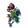
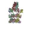
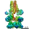
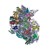
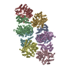
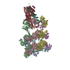
| Title | Human nuclear RNA exosome EXO-14 complex | |||||||||
 Map data Map data | Cryo-EM reconstruction of the human nuclear RNA exosome EXO-14 complex | |||||||||
 Sample Sample |
| |||||||||
| Biological species | synthetic construct (others) /  Homo sapiens (human) Homo sapiens (human) | |||||||||
| Method | single particle reconstruction / cryo EM / Resolution: 6.25 Å | |||||||||
 Authors Authors | Gerlach P / Schuller JM / Falk S / Basquin J / Conti E | |||||||||
 Citation Citation | Journal: Elife / Year: 2018 Title: Distinct and evolutionary conserved structural features of the human nuclear exosome complex. Authors: Piotr Gerlach / Jan M Schuller / Fabien Bonneau / Jérôme Basquin / Peter Reichelt / Sebastian Falk / Elena Conti /  Abstract: The nuclear RNA exosome complex mediates the processing of structured RNAs and the decay of aberrant non-coding RNAs, an important function particularly in human cells. Most mechanistic studies to ...The nuclear RNA exosome complex mediates the processing of structured RNAs and the decay of aberrant non-coding RNAs, an important function particularly in human cells. Most mechanistic studies to date have focused on the yeast system. Here, we reconstituted and studied the properties of a recombinant 14-subunit human nuclear exosome complex. In biochemical assays, the human exosome embeds a longer RNA channel than its yeast counterpart. The 3.8 Å resolution cryo-EM structure of the core complex bound to a single-stranded RNA reveals that the RNA channel path is formed by two distinct features of the hDIS3 exoribonuclease: an open conformation and a domain organization more similar to bacterial RNase II than to yeast Rrp44. The cryo-EM structure of the holo-complex shows how obligate nuclear cofactors position the hMTR4 helicase at the entrance of the core complex, suggesting a striking structural conservation from lower to higher eukaryotes. | |||||||||
| History |
|
- Structure visualization
| Movie |
 Movie viewer Movie viewer |
|---|---|
| Structure viewer | EM map: SurfViewMolmilJmol/JSmol |
| Supplemental images |

- Downloads & links
-EMDB archive
| Map data | emd_0127.map.gz | 5.4 MB | EMDB map data format | |
|---|---|---|---|---|
| Header (meta data) | emd-0127-v30.xmlemd-0127.xml | 28.6 KB 28.6 KB | Display Display | EMDB header |
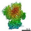
| Images | 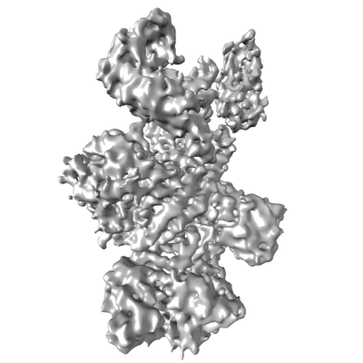 emd_0127.png emd_0127.png | 55.2 KB | ||
| Archive directory |  http://ftp.pdbj.org/pub/emdb/structures/EMD-0127ftp://ftp.pdbj.org/pub/emdb/structures/EMD-0127 http://ftp.pdbj.org/pub/emdb/structures/EMD-0127ftp://ftp.pdbj.org/pub/emdb/structures/EMD-0127 | HTTPS FTP |
-Related structure data


-Links
| EMDB pages | EMDB (EBI/PDBe) / EMDataResource |
|---|
-Map
| File | Download / File: emd_0127.map.gz / Format: CCP4 / Size: 59.6 MB / Type: IMAGE STORED AS FLOATING POINT NUMBER (4 BYTES) | ||||||||||||||||||||||||||||||||||||||||||||||||||||||||||||
|---|---|---|---|---|---|---|---|---|---|---|---|---|---|---|---|---|---|---|---|---|---|---|---|---|---|---|---|---|---|---|---|---|---|---|---|---|---|---|---|---|---|---|---|---|---|---|---|---|---|---|---|---|---|---|---|---|---|---|---|---|---|
| Annotation | Cryo-EM reconstruction of the human nuclear RNA exosome EXO-14 complex | ||||||||||||||||||||||||||||||||||||||||||||||||||||||||||||
| Projections & slices | Image control
Images are generated by Spider. | ||||||||||||||||||||||||||||||||||||||||||||||||||||||||||||
| Voxel size | X=Y=Z: 1.35 Å | ||||||||||||||||||||||||||||||||||||||||||||||||||||||||||||
| Density |
| ||||||||||||||||||||||||||||||||||||||||||||||||||||||||||||
| Symmetry | Space group: 1 | ||||||||||||||||||||||||||||||||||||||||||||||||||||||||||||
| Details | EMDB XML:
CCP4 map header:
| ||||||||||||||||||||||||||||||||||||||||||||||||||||||||||||
 Z (Sec.)
 Y (Row.)
 X (Col.)


-Supplemental data
- Sample components
+Entire : Human nuclear RNA exosome EXO-14 complex
+Supramolecule #1: Human nuclear RNA exosome EXO-14 complex
+Supramolecule #2: Human nuclear RNA exosome EXO-14 complex

+Supramolecule #3: Human nuclear RNA exosome EXO-14 complex
+Macromolecule #1: EXOSC9
+Macromolecule #2: EXOSC4
+Macromolecule #3: EXOSC8
+Macromolecule #4: EXOSC5
+Macromolecule #5: EXOSC7
+Macromolecule #6: EXOSC6
+Macromolecule #7: EXOSC3
+Macromolecule #8: EXOSC2
+Macromolecule #9: EXOSC1
+Macromolecule #10: DIS3
+Macromolecule #11: MPP6
+Macromolecule #12: EXOSC10
+Macromolecule #13: C1D
+Macromolecule #14: MTR4
+Macromolecule #15: U44 ssRNA
-Experimental details
-Structure determination
| Method | cryo EM |
|---|---|
 Processing Processing | single particle reconstruction |
| Aggregation state | particle |
-Sample preparation
| Concentration | 0.3 mg/mL | ||||||||
|---|---|---|---|---|---|---|---|---|---|
| Buffer | pH: 7.5 Component:
| ||||||||
| Vitrification | Cryogen name: ETHANE-PROPANE / Instrument: FEI VITROBOT MARK IV |
- Electron microscopy
| Microscope | FEI TITAN KRIOS |
|---|---|
| Image recording | Film or detector model: GATAN K2 SUMMIT (4k x 4k) / Detector mode: COUNTING / Number real images: 8047 / Average electron dose: 47.0 e/Å2 |
| Electron beam | Acceleration voltage: 300 kV / Electron source:  FIELD EMISSION GUN FIELD EMISSION GUN |
| Electron optics | Illumination mode: FLOOD BEAM / Imaging mode: BRIGHT FIELD |
| Experimental equipment |  Model: Titan Krios / Image courtesy: FEI Company |
