 Movie
Movie Controller


+ Open data

- Basic information
| Entry |  | |||||||||
|---|---|---|---|---|---|---|---|---|---|---|
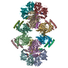
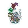
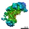
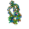
| Title | GAPDH-CP12-PRK complex | |||||||||
 Map data Map data | None | |||||||||
 Sample Sample |
| |||||||||
 Keywords Keywords | complex / calvin cycle / redox regulation / PHOTOSYNTHESIS | |||||||||
| Function / homology |  Function and homology information Function and homology informationphosphoribulokinase / phosphoribulokinase activity / negative regulation of reductive pentose-phosphate cycle / glyceraldehyde-3-phosphate dehydrogenase (NADP+) (phosphorylating) activity / Oxidoreductases; Acting on the aldehyde or oxo group of donors; With NAD+ or NADP+ as acceptor / reductive pentose-phosphate cycle / glyceraldehyde-3-phosphate dehydrogenase (NAD+) (phosphorylating) activity / glucose metabolic process / NAD binding / NADP binding ...phosphoribulokinase / phosphoribulokinase activity / negative regulation of reductive pentose-phosphate cycle / glyceraldehyde-3-phosphate dehydrogenase (NADP+) (phosphorylating) activity / Oxidoreductases; Acting on the aldehyde or oxo group of donors; With NAD+ or NADP+ as acceptor / reductive pentose-phosphate cycle / glyceraldehyde-3-phosphate dehydrogenase (NAD+) (phosphorylating) activity / glucose metabolic process / NAD binding / NADP binding / ATP binding / metal ion binding Similarity search - Function | |||||||||
| Biological species |   Thermosynechococcus elongatus BP-1 (bacteria) / Thermosynechococcus elongatus (strain BP-1) (bacteria) Thermosynechococcus elongatus BP-1 (bacteria) / Thermosynechococcus elongatus (strain BP-1) (bacteria) | |||||||||
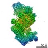
| Method | single particle reconstruction / cryo EM / Resolution: 3.9 Å | |||||||||
 Authors Authors | McFarlane CR / Shah N | |||||||||
| Funding support |  United Kingdom, 1 items United Kingdom, 1 items
| |||||||||
 Citation Citation | Journal: Proc Natl Acad Sci U S A / Year: 2019 Title: Structural basis of light-induced redox regulation in the Calvin-Benson cycle in cyanobacteria. Authors: Ciaran R McFarlane / Nita R Shah / Burak V Kabasakal / Blanca Echeverria / Charles A R Cotton / Doryen Bubeck / James W Murray / Abstract: Plants, algae, and cyanobacteria fix carbon dioxide to organic carbon with the Calvin-Benson (CB) cycle. Phosphoribulokinase (PRK) and glyceraldehyde 3-phosphate dehydrogenase (GAPDH) are essential ...Plants, algae, and cyanobacteria fix carbon dioxide to organic carbon with the Calvin-Benson (CB) cycle. Phosphoribulokinase (PRK) and glyceraldehyde 3-phosphate dehydrogenase (GAPDH) are essential CB-cycle enzymes that control substrate availability for the carboxylation enzyme Rubisco. PRK consumes ATP to produce the Rubisco substrate ribulose bisphosphate (RuBP). GAPDH catalyzes the reduction step of the CB cycle with NADPH to produce the sugar glyceraldehyde 3-phosphate (GAP), which is used for regeneration of RuBP and is the main exit point of the cycle. GAPDH and PRK are coregulated by the redox state of a conditionally disordered protein CP12, which forms a ternary complex with both enzymes. However, the structural basis of CB-cycle regulation by CP12 is unknown. Here, we show how CP12 modulates the activity of both GAPDH and PRK. Using thermophilic cyanobacterial homologs, we solve crystal structures of GAPDH with different cofactors and CP12 bound, and the ternary GAPDH-CP12-PRK complex by electron cryo-microscopy, we reveal that formation of the N-terminal disulfide preorders CP12 prior to binding the PRK active site, which is resolved in complex with CP12. We find that CP12 binding to GAPDH influences substrate accessibility of all GAPDH active sites in the binary and ternary inhibited complexes. Our structural and biochemical data explain how CP12 integrates responses from both redox state and nicotinamide dinucleotide availability to regulate carbon fixation. | |||||||||
| History |
|
- Structure visualization
| Structure viewer | EM map: SurfViewMolmilJmol/JSmol |
|---|---|
| Supplemental images |

- Downloads & links
-EMDB archive
| Map data | emd_0071.map.gz | 152.4 MB | EMDB map data format | |
|---|---|---|---|---|
| Header (meta data) | emd-0071-v30.xmlemd-0071.xml | 20.8 KB 20.8 KB | Display Display | EMDB header |
| FSC (resolution estimation) | emd_0071_fsc.xml | 12.5 KB | Display | FSC data file |
| Images | 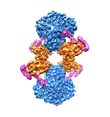 emd_0071.png emd_0071.png | 118 KB | ||
| Filedesc metadata | emd-0071.cif.gz | 6.9 KB | ||
| Others | emd_0071_half_map_1.map.gzemd_0071_half_map_2.map.gz | 129 MB 129 MB | ||
| Archive directory |  http://ftp.pdbj.org/pub/emdb/structures/EMD-0071ftp://ftp.pdbj.org/pub/emdb/structures/EMD-0071 http://ftp.pdbj.org/pub/emdb/structures/EMD-0071ftp://ftp.pdbj.org/pub/emdb/structures/EMD-0071 | HTTPS FTP |
-Related structure data
| Related structure data |  6gveMC  6gfoC  6gfpC  6gfqC  6gfrC 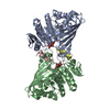 6gg7C  6ghlC 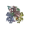 6ghrC M: atomic model generated by this map C: citing same article ( |
|---|---|
| Similar structure data |


-Links
| EMDB pages | EMDB (EBI/PDBe) / EMDataResource |
|---|---|
| Related items in Molecule of the Month |

-Map
| File | Download / File: emd_0071.map.gz / Format: CCP4 / Size: 163.6 MB / Type: IMAGE STORED AS FLOATING POINT NUMBER (4 BYTES) | ||||||||||||||||||||||||||||||||||||
|---|---|---|---|---|---|---|---|---|---|---|---|---|---|---|---|---|---|---|---|---|---|---|---|---|---|---|---|---|---|---|---|---|---|---|---|---|---|
| Annotation | None | ||||||||||||||||||||||||||||||||||||
| Projections & slices | Image control
Images are generated by Spider. | ||||||||||||||||||||||||||||||||||||
| Voxel size | X=Y=Z: 1.047 Å | ||||||||||||||||||||||||||||||||||||
| Density |
| ||||||||||||||||||||||||||||||||||||
| Symmetry | Space group: 1 | ||||||||||||||||||||||||||||||||||||
| Details | EMDB XML:
|
 Z (Sec.)
 Y (Row.)
 X (Col.)


-Supplemental data
-Half map: half map 1
| File | emd_0071_half_map_1.map | ||||||||||||
|---|---|---|---|---|---|---|---|---|---|---|---|---|---|
| Annotation | half map 1 | ||||||||||||
| Projections & Slices |
| ||||||||||||
| Density Histograms |


-Half map: half map 2
| File | emd_0071_half_map_2.map | ||||||||||||
|---|---|---|---|---|---|---|---|---|---|---|---|---|---|
| Annotation | half map 2 | ||||||||||||
| Projections & Slices |
| ||||||||||||
| Density Histograms |


- Sample components
-Entire : GAPDH-CP12-PRK complex
| Entire | Name: GAPDH-CP12-PRK complex |
|---|---|
| Components |
|
-Supramolecule #1: GAPDH-CP12-PRK complex
| Supramolecule | Name: GAPDH-CP12-PRK complex / type: complex / ID: 1 / Parent: 0 / Macromolecule list: #1-#3 |
|---|---|
| Molecular weight | Theoretical: 480 KDa |
-Supramolecule #2: GAPDH-CP12-PRK complex
| Supramolecule | Name: GAPDH-CP12-PRK complex / type: complex / ID: 2 / Parent: 1 / Macromolecule list: #1 |
|---|---|
| Source (natural) | Organism: Thermosynechococcus elongatus BP-1 (bacteria) |
-Supramolecule #3: GAPDH-CP12-PRK complex
| Supramolecule | Name: GAPDH-CP12-PRK complex / type: complex / ID: 3 / Parent: 1 / Macromolecule list: #2-#3 |
|---|---|
| Source (natural) | Organism: Thermosynechococcus elongatus BP-1 (bacteria) |
-Macromolecule #1: CP12 polypeptide
| Macromolecule | Name: CP12 polypeptide / type: protein_or_peptide / ID: 1 / Number of copies: 4 / Enantiomer: LEVO |
|---|---|
| Source (natural) | Organism: Thermosynechococcus elongatus (strain BP-1) (bacteria) Strain: BP-1 |
| Molecular weight | Theoretical: 8.611127 KDa |
| Recombinant expression | Organism: |
| Sequence | String: GSMSNLEKQI EQAREEAHKI CDTEGATSGQ CAAAWDALEE LQAEAAHQRA EQQDHKTSFQ QYCDDNPDAA ECRIYDD UniProtKB: CP12 polypeptide |
-Macromolecule #2: Glyceraldehyde-3-phosphate dehydrogenase
| Macromolecule | Name: Glyceraldehyde-3-phosphate dehydrogenase / type: protein_or_peptide / ID: 2 / Number of copies: 8 / Enantiomer: LEVO EC number: Oxidoreductases; Acting on the aldehyde or oxo group of donors; With NAD+ or NADP+ as acceptor |
|---|---|
| Source (natural) | Organism: Thermosynechococcus elongatus (strain BP-1) (bacteria) Strain: BP-1 |
| Molecular weight | Theoretical: 36.792734 KDa |
| Recombinant expression | Organism: |
| Sequence | String: GSMVRVAING FGRIGRNFMR CWLQRKANSK LEIVGINDTS DPRTNAHLLK YDSMLGIFQD AEITADDDCI YAGGHAVKCV SDRNPENLP WSAWGIDLVI EATGVFTSRE GASKHLSAGA KKVLITAPGK GNIPTYVVGV NHHTYDPSED IVSNASCTTN C LAPIVKVL ...String: GSMVRVAING FGRIGRNFMR CWLQRKANSK LEIVGINDTS DPRTNAHLLK YDSMLGIFQD AEITADDDCI YAGGHAVKCV SDRNPENLP WSAWGIDLVI EATGVFTSRE GASKHLSAGA KKVLITAPGK GNIPTYVVGV NHHTYDPSED IVSNASCTTN C LAPIVKVL HEAFGIQQGM MTTTHSYTGD QRLLDASHRD LRRARAAAMN IVPTSTGAAK AVGLVIPELQ GKLNGIALRV PT PNVSVVD FVAQVEKPTI AEQVNQVIKE ASETTMKGII HYSELELVSS DYRGHNASSI LDASLTMVLG GNLVKVVAWY DNE WGYSQR VLDLAEHMAA HWA UniProtKB: Glyceraldehyde-3-phosphate dehydrogenase |
-Macromolecule #3: Phosphoribulokinase
| Macromolecule | Name: Phosphoribulokinase / type: protein_or_peptide / ID: 3 / Number of copies: 4 / Enantiomer: LEVO / EC number: phosphoribulokinase |
|---|---|
| Source (natural) | Organism: Thermosynechococcus elongatus (strain BP-1) (bacteria) Strain: BP-1 |
| Molecular weight | Theoretical: 38.038234 KDa |
| Sequence | String: MSSKPDRVVL IGVAGDSGCG KSTFLRRLAD LFGEDFMTVI CLDDYHSLDR KQRKEMGITA LDPRANNFDL MYEQIKALKN GESIMKPIY NHETGTIDPP EKVDPNHVIV IEGLHPLYDE RVRSLIDFSV YLDISDDVKI AWKIKRDMAE RGHSYEDVIA S INARRPDF ...String: MSSKPDRVVL IGVAGDSGCG KSTFLRRLAD LFGEDFMTVI CLDDYHSLDR KQRKEMGITA LDPRANNFDL MYEQIKALKN GESIMKPIY NHETGTIDPP EKVDPNHVIV IEGLHPLYDE RVRSLIDFSV YLDISDDVKI AWKIKRDMAE RGHSYEDVIA S INARRPDF MAYIDPQKQY ADVVLQILPS QLAKEEKVGN ILRVRMLQRE GIPGFEPVYL FDEGSTITWI PCGRKLTCSY PG IRLSYGP DEYYGHPVSV LEVDGRFEKL DELIYIESHL SNTSTKHYGE VTELLLKHRD YPGSDNGSGL FQVLTGLKMR ATY ERLTSR DAATVTNR UniProtKB: Phosphoribulokinase |
-Macromolecule #4: NICOTINAMIDE-ADENINE-DINUCLEOTIDE
| Macromolecule | Name: NICOTINAMIDE-ADENINE-DINUCLEOTIDE / type: ligand / ID: 4 / Number of copies: 8 / Formula: NAD |
|---|---|
| Molecular weight | Theoretical: 663.425 Da |
| Chemical component information |  ChemComp-NAD: |
-Experimental details
-Structure determination
| Method | cryo EM |
|---|---|
 Processing Processing | single particle reconstruction |
| Aggregation state | particle |
-Sample preparation
| Buffer | pH: 7.9 Component:
| |||||||||||||||||||||
|---|---|---|---|---|---|---|---|---|---|---|---|---|---|---|---|---|---|---|---|---|---|---|
| Grid | Model: Quantifoil R2/2 / Material: COPPER / Mesh: 300 / Support film - Material: CARBON / Support film - topology: CONTINUOUS / Support film - Film thickness: 3 / Pretreatment - Type: GLOW DISCHARGE / Pretreatment - Time: 40 sec. / Pretreatment - Atmosphere: AIR | |||||||||||||||||||||
| Vitrification | Cryogen name: ETHANE / Chamber humidity: 85 % / Chamber temperature: 283 K / Instrument: FEI VITROBOT MARK IV Details: Blot force 3. Wait time 60 seconds, then blotted for 3.5 seconds before plunging. 2.5 ul of sample.. |
- Electron microscopy
| Microscope | FEI TITAN KRIOS |
|---|---|
| Image recording | Film or detector model: GATAN K2 SUMMIT (4k x 4k) / Detector mode: COUNTING / Average electron dose: 44.0 e/Å2 |
| Electron beam | Acceleration voltage: 300 kV / Electron source:  FIELD EMISSION GUN FIELD EMISSION GUN |
| Electron optics | Illumination mode: FLOOD BEAM / Imaging mode: BRIGHT FIELD |
| Sample stage | Specimen holder model: FEI TITAN KRIOS AUTOGRID HOLDER / Cooling holder cryogen: NITROGEN |
| Experimental equipment |  Model: Titan Krios / Image courtesy: FEI Company |
+Image processing

-Atomic model buiding 1
| Refinement | Space: REAL / Protocol: FLEXIBLE FIT |
|---|---|
| Output model | PDB-6gve: |
