Movie
Movie Controller

[English] 日本語
 Yorodumi
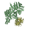
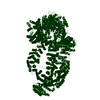
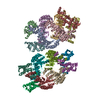
- PDB-5oej: Structure of Tra1 subunit within the chromatin modifying complex SAGA -
+ Open data

- Basic information
| Entry | Database: PDB / ID: 5oej | |||||||||
|---|---|---|---|---|---|---|---|---|---|---|
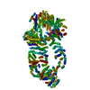
| Title | Structure of Tra1 subunit within the chromatin modifying complex SAGA | |||||||||
 Components Components | Tra1 subunit within the chromatin modifying complex SAGA | |||||||||
 Keywords Keywords | TRANSCRIPTION / yeast / Tra1 / SAGA / activator target | |||||||||
| Function / homology |  Function and homology information Function and homology informationSLIK (SAGA-like) complex / SAGA complex / histone acetyltransferase activity / NuA4 histone acetyltransferase complex / transferase activity / DNA repair / regulation of DNA-templated transcription / positive regulation of transcription by RNA polymerase II / nucleus Similarity search - Function | |||||||||
| Biological species |  Komagataella pastoris (fungus) Komagataella pastoris (fungus) | |||||||||
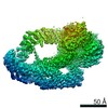
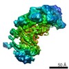
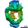
| Method | ELECTRON MICROSCOPY / single particle reconstruction / cryo EM / Resolution: 5.7 Å | |||||||||
 Authors Authors | Sharov, G. / Voltz, K. / Durand, A. / Kolesnikova, O. / Papai, G. / Myasnikov, A.G. / Dejaegere, A. / Ben-Shem, A. / Schultz, P. | |||||||||
| Funding support |  France, 2items France, 2items
| |||||||||
 Citation Citation | Journal: Nat Commun / Year: 2017 Title: Structure of the transcription activator target Tra1 within the chromatin modifying complex SAGA. Authors: Grigory Sharov / Karine Voltz / Alexandre Durand / Olga Kolesnikova / Gabor Papai / Alexander G Myasnikov / Annick Dejaegere / Adam Ben Shem / Patrick Schultz /   Abstract: The transcription co-activator complex SAGA is recruited to gene promoters by sequence-specific transcriptional activators and by chromatin modifications to promote pre-initiation complex formation. ...The transcription co-activator complex SAGA is recruited to gene promoters by sequence-specific transcriptional activators and by chromatin modifications to promote pre-initiation complex formation. The yeast Tra1 subunit is the major target of acidic activators such as Gal4, VP16, or Gcn4 but little is known about its structural organization. The 430 kDa Tra1 subunit and its human homolog the transformation/transcription domain-associated protein TRRAP are members of the phosphatidyl 3-kinase-related kinase (PIKK) family. Here, we present the cryo-EM structure of the entire SAGA complex where the major target of activator binding, the 430 kDa Tra1 protein, is resolved with an average resolution of 5.7 Å. The high content of alpha-helices in Tra1 enabled tracing of the majority of its main chain. Our results highlight the integration of Tra1 within the major epigenetic regulator SAGA. | |||||||||
| History |
|
- Structure visualization
| Movie |
Movie viewer |
|---|---|
| Structure viewer | Molecule: MolmilJmol/JSmol |
- Downloads & links
-Download
| PDBx/mmCIF format | 5oej.cif.gz | 420.6 KB | Display | PDBx/mmCIF format |
|---|---|---|---|---|
| PDB format | pdb5oej.ent.gz | 268.3 KB | Display | PDB format |
| PDBx/mmJSON format | 5oej.json.gz | Tree view | PDBx/mmJSON format | |
| Others |  Other downloads Other downloads |
-Validation report
| Arichive directory | https://data.pdbj.org/pub/pdb/validation_reports/oe/5oejftp://data.pdbj.org/pub/pdb/validation_reports/oe/5oej | HTTPS FTP |
|---|
-Related structure data
| Related structure data | 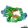 3790MC 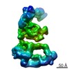 3804C M: map data used to model this data C: citing same article ( |
|---|---|
| Similar structure data |

-Links
 PDBj
PDBj
- Assembly
| Deposited unit | 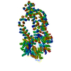
|
|---|---|
| 1 |
|
-Components
| #1: Protein | Mass: 438055.344 Da / Num. of mol.: 1 / Source method: isolated from a natural source / Source: (natural) Komagataella pastoris (fungus) / References: UniProt: F2QQ15, UniProt: C4QYV4*PLUS |
|---|
-Experimental details
-Experiment
| Experiment | Method: ELECTRON MICROSCOPY |
|---|---|
| EM experiment | Aggregation state: PARTICLE / 3D reconstruction method: single particle reconstruction |
- Sample preparation
| Component | Name: Tra1 subunit of SAGA complex / Type: COMPLEX / Entity ID: all / Source: NATURAL |
|---|---|
| Molecular weight | Value: 0.4 MDa / Experimental value: NO |
| Source (natural) | Organism: Komagataella pastoris (fungus) |
| Buffer solution | pH: 8 |
| Specimen | Conc.: 0.2 mg/ml / Embedding applied: NO / Shadowing applied: NO / Staining applied: NO / Vitrification applied: YES |
| Specimen support | Grid material: COPPER / Grid mesh size: 300 divisions/in. / Grid type: Quantifoil R2/2 |
| Vitrification | Instrument: FEI VITROBOT MARK IV / Cryogen name: ETHANE / Humidity: 95 % / Chamber temperature: 277 K / Details: Blot for 1 second before plunging |
- Electron microscopy imaging
| Experimental equipment |  Model: Titan Krios / Image courtesy: FEI Company |
|---|---|
| Microscopy | Model: FEI TITAN KRIOS |
| Electron gun | Electron source:  FIELD EMISSION GUN / Accelerating voltage: 300 kV / Illumination mode: FLOOD BEAM FIELD EMISSION GUN / Accelerating voltage: 300 kV / Illumination mode: FLOOD BEAM |
| Electron lens | Mode: BRIGHT FIELD / Nominal magnification: 59000 X / Calibrated magnification: 127272 X / Nominal defocus max: 3400 nm / Nominal defocus min: 1400 nm / Calibrated defocus min: 1400 nm / Calibrated defocus max: 3400 nm / Cs: 0.001 mm / C2 aperture diameter: 100 µm / Alignment procedure: ZEMLIN TABLEAU |
| Specimen holder | Cryogen: NITROGEN / Specimen holder model: FEI TITAN KRIOS AUTOGRID HOLDER / Temperature (max): 80 K / Temperature (min): 70 K |
| Image recording | Average exposure time: 1 sec. / Electron dose: 60 e/Å2 / Detector mode: INTEGRATING / Film or detector model: FEI FALCON II (4k x 4k) / Num. of grids imaged: 4 / Num. of real images: 8505 Details: Images were collected in movie-mode at 17 frames per second, frame 1 was not acquired. Every two frames were joined together, producing 8 frames per second. |
| EM imaging optics | Spherical aberration corrector: Microscope has a Cs corrector |
| Image scans | Sampling size: 14 µm / Width: 4096 / Height: 4096 / Movie frames/image: 8 / Used frames/image: 2-8 |
- Processing
| EM software |
| |||||||||||||||||||||||||||||||||||||||||||||||||||||||
|---|---|---|---|---|---|---|---|---|---|---|---|---|---|---|---|---|---|---|---|---|---|---|---|---|---|---|---|---|---|---|---|---|---|---|---|---|---|---|---|---|---|---|---|---|---|---|---|---|---|---|---|---|---|---|---|---|
| CTF correction | Details: Full CTF correction in Relion / Type: PHASE FLIPPING AND AMPLITUDE CORRECTION | |||||||||||||||||||||||||||||||||||||||||||||||||||||||
| Particle selection | Num. of particles selected: 264901 | |||||||||||||||||||||||||||||||||||||||||||||||||||||||
| Symmetry | Point symmetry: C1 (asymmetric) | |||||||||||||||||||||||||||||||||||||||||||||||||||||||
| 3D reconstruction | Resolution: 5.7 Å / Resolution method: FSC 0.143 CUT-OFF / Num. of particles: 105916 / Algorithm: FOURIER SPACE / Symmetry type: POINT | |||||||||||||||||||||||||||||||||||||||||||||||||||||||
| Atomic model building | Protocol: RIGID BODY FIT / Space: REAL Details: Secondary structure restraints were applied in Phenix. | |||||||||||||||||||||||||||||||||||||||||||||||||||||||
| Atomic model building | PDB-ID: 4JSN Pdb chain-ID: B / Accession code: 4JSN / Pdb chain residue range: 1385-2549 / Source name: PDB / Type: experimental model | |||||||||||||||||||||||||||||||||||||||||||||||||||||||
| Refinement | Highest resolution: 5.7 Å |