 Movie
Movie Controller
[English] 日本語
 Yorodumi
- SASDBY4: Pentameric Nucleoplasmin-histone H2A/H2B complex (Nucleoplasmin c... -
+ Open data

- Basic information
| Entry | Database: SASBDB / ID: SASDBY4 |
|---|---|
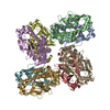
 Sample Sample | Pentameric Nucleoplasmin-histone H2A/H2B complex
|
| Function / homology |  Function and homology information Function and homology informationstructural constituent of chromatin / nucleosome / heterochromatin formation / protein heterodimerization activity / DNA binding / nucleus Similarity search - Function |
| Biological species | |
 Citation Citation | Date: 2017 Dec Title: Dynamic intramolecular regulation of the histone chaperone nucleoplasmin controls histone binding and release Authors: Warren C / Matsui T / Karp J / Onikubo T / Cahill S / Brenowitz M / Cowburn D / Girvin M |
 Contact author Contact author |
|
- Structure visualization
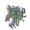
| Structure viewer | Molecule: MolmilJmol/JSmol |
|---|
- Downloads & links
-Data source
| SASBDB page |  SASDBY4 SASDBY4 |
|---|
-Related structure data
| Similar structure data |
|---|


-External links
| Related items in Molecule of the Month |
|---|


-Models
| Model #546 |  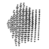 Type: dummy / Software: (r6345) / Radius of dummy atoms: 5.75 A / Symmetry: P5 / Chi-square value: 1.580  Search similar-shape structures of this assembly by Omokage search (details) Search similar-shape structures of this assembly by Omokage search (details) |
|---|---|
| Model #1701 |   Type: mix / Radius of dummy atoms: 1.90 A / Symmetry: P5 / Chi-square value: 1.43 Search similar-shape structures of this assembly by Omokage search (details) |
| Model #1702 |  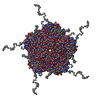 Type: mix / Radius of dummy atoms: 1.90 A / Symmetry: P5 / Chi-square value: 1.26 / P-value: 0.000139 Search similar-shape structures of this assembly by Omokage search (details) |
-Sample
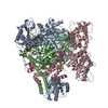
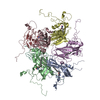
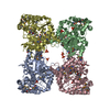
| Sample | Name: Pentameric Nucleoplasmin-histone H2A/H2B complex / Entity id: 348 / 349 / 350 |
|---|---|
| Buffer | Name: 20 mM Tris 150mM NaCl 1mM EDTA 5mM DTT / Concentration: 20.00 mM / pH: 8 / Composition: 150mM NaCl, 1mM EDTA, 5mM DTT |
| Entity #348 | Name: Npm core / Type: protein / Description: Nucleoplasmin core + A2 / Formula weight: 16.165 / Num. of mol.: 5 / Source: Xenopus laevis / References: UniProt: Q6NUC7 Sequence: ASTASNTSKV EKPVSLIWGC ELNEQNKTFA FKIEDEEEKC EHQLALRTVC LGDKAKDEFH IVEIVTQEEG KEKPVPIASL KPSILPMATM VGIELTPPVT FRLKAGSGPV YISGQHVAME EDYSWAEEED EGEEEEEEEE DPESP |
| Entity #349 | Name: 2HA / Type: protein / Description: Histone H2A (ΔAla127) / Formula weight: 13.879 / Num. of mol.: 5 / Source: Xenopus laevis / References: UniProt: Q6AZJ8 Sequence: SGRGKQGGKT RAKAKTRSSR AGLQFPVGRV HRLLRKGNYA ERVGAGAPVY LAAVLEYLTA EILELAGNAA RDNKKTRIIP RHLQLAVRND EELNKLLGRV TIAQGGVLPN IQSVLLPKKT ESSKSKSK |
| Entity #350 | Name: H2B / Type: protein / Description: Histone H2B 1.1 (Ser33Thr) / Formula weight: 13.494 / Num. of mol.: 5 / Source: Xenopus laevis / References: UniProt: P02281 Sequence: AKSAPAPKKG SKKAVTKTQK KDGKKRRKTR KESYAIYVYK VLKQVHPDTG ISSKAMSIMN SFVNDVFERI AGEASRLAHY NKRSTITSRE IQTAVRLLLP GELAKHAVSE GTKAVTKYTS AK |
-Experimental information
| Beam | Instrument name: Stanford Synchrotron Radiation Lightsource (SSRL) BL4-2 City: Stanford, CA / 国: USA  / Type of source: X-ray synchrotron / Wavelength: 0.1127 Å / Dist. spec. to detc.: 1.7 mm / Type of source: X-ray synchrotron / Wavelength: 0.1127 Å / Dist. spec. to detc.: 1.7 mm | ||||||||||||||||||||||||||||||||||||
|---|---|---|---|---|---|---|---|---|---|---|---|---|---|---|---|---|---|---|---|---|---|---|---|---|---|---|---|---|---|---|---|---|---|---|---|---|---|
| Detector | Name: Rayonix MX225-HE | ||||||||||||||||||||||||||||||||||||
| Scan | 
| ||||||||||||||||||||||||||||||||||||
| Distance distribution function P(R) | 
| ||||||||||||||||||||||||||||||||||||
| Result |  Comments: The models displayed for this entry are: Top: ab initio bead model representation (derived from individual reconstructions (example fit shown), aligned, spatially averaged and volume- ...Comments: The models displayed for this entry are: Top: ab initio bead model representation (derived from individual reconstructions (example fit shown), aligned, spatially averaged and volume-occupancy corrected): Middle: The best CORAL model using the major A2 site (site #1) of A2:H2A/H2B complex as a starting structure (See Figure 5D in the primary citation). Lacking fragments reconstructed by the program CORAL are shown as spheres. Bottom: The best CORAL model using the minor A2 site (site #2) of A2:H2A/H2B complex as a starting structure (See Figure 5D in the primary citation). Lacking fragments reconstructed by the program CORAL are shown as spheres.
|
