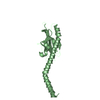
Journal: Anal Biochem / Year: 2009 Title: Characterization of a fluorophore binding RNA aptamer by fluorescence correlation spectroscopy and small angle X-ray scattering. Authors: Arne Werner / Petr V Konarev / Dmitri I Svergun / Ulrich Hahn / Abstract: Using fluorescence correlation spectroscopy (FCS), we have established an in vitro assay to study RNA dynamics by analyzing fluorophore binding RNA aptamers at the single molecule level. The RNA ...Using fluorescence correlation spectroscopy (FCS), we have established an in vitro assay to study RNA dynamics by analyzing fluorophore binding RNA aptamers at the single molecule level. The RNA aptamer SRB2m, a minimized variant of the initially selected aptamer SRB-2, has a high affinity to the disulfonated triphenylmethane dye sulforhodamine B. A mobility shift of sulforhodamine B after binding to SRB2m was measured. In contrast, patent blue V (PBV) is visible only if complexed with SRB2m due to increased molecular brightness and minimal background. With small angle X-ray scattering (SAXS), the three-dimensional structure of the RNA aptamer was characterized at low resolution to analyze the effect of fluorophore binding. The aptamer and sulforhodamine B-aptamer complex was found to be predominantly dimeric in solution. Interaction of PBV with SRB2m led to a dissociation of SRB2m dimers into monomers. Radii of gyration and hydrodynamic radii, gained from dynamic light scattering, FCS, and fluorescence cross-correlation experiments, led to comparable conclusions. Our study demonstrates how RNA-aptamer fluorophore complexes can be simultaneously structurally and photophysically characterized by FCS. Furthermore, fluorophore binding RNA aptamers provide a tool for visualizing single RNA molecules.
Contact author
Petr Konarev (EMBL-Hamburg, European Molecular Biology Laboratory (EMBL) - Hamburg outstation, Notkestraße 85, Geb. 25A, 22607 Hamburg, Deutschland, Germany)
Instrument name: DORIS III X33 / City: Hamburg / 国: Germany / Type of source: X-ray synchrotron
Detector
Name: MAR 345 Image Plate
Scan
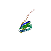
Title: SRB2m with PBV / Measurement date: Oct 24, 2007 / Storage temperature: 15 °C / Cell temperature: 15 °C / Exposure time: 120 sec. / Number of frames: 2 / Unit: 1/nm /
Min
Max
Q
0.2449
5.0717
Distance distribution function P(R)
Sofotware P(R): GNOM 4.5a / Number of points: 512 /
Min
Max
Q
0.2483
4.98
P(R) point
1
512
R
0
8
Result
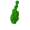
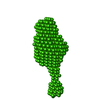
D max: 8 / Type of curve: single_conc /
Experimental
Porod
MW
20 kDa
-
Volume
-
24 nm3
P(R)
Guinier
Forward scattering, I0
177
180.23
Radius of gyration, Rg
2.27 nm
2.25 nm
+
About Yorodumi
-
News
-
Feb 9, 2022. New format data for meta-information of EMDB entries
New format data for meta-information of EMDB entries
Version 3 of the EMDB header file is now the official format.
The previous official version 1.9 will be removed from the archive.
In the structure databanks used in Yorodumi, some data are registered as the other names, "COVID-19 virus" and "2019-nCoV". Here are the details of the virus and the list of structure data.
Jan 31, 2019. EMDB accession codes are about to change! (news from PDBe EMDB page)
EMDB accession codes are about to change! (news from PDBe EMDB page)
The allocation of 4 digits for EMDB accession codes will soon come to an end. Whilst these codes will remain in use, new EMDB accession codes will include an additional digit and will expand incrementally as the available range of codes is exhausted. The current 4-digit format prefixed with “EMD-” (i.e. EMD-XXXX) will advance to a 5-digit format (i.e. EMD-XXXXX), and so on. It is currently estimated that the 4-digit codes will be depleted around Spring 2019, at which point the 5-digit format will come into force.
The EM Navigator/Yorodumi systems omit the EMD- prefix.
Related info.:Q: What is EMD? / ID/Accession-code notation in Yorodumi/EM Navigator
Yorodumi is a browser for structure data from EMDB, PDB, SASBDB, etc.
This page is also the successor to EM Navigator detail page, and also detail information page/front-end page for Omokage search.
The word "yorodu" (or yorozu) is an old Japanese word meaning "ten thousand". "mi" (miru) is to see.
Related info.:EMDB / PDB / SASBDB / Comparison of 3 databanks / Yorodumi Search / Aug 31, 2016. New EM Navigator & Yorodumi / Yorodumi Papers / Jmol/JSmol / Function and homology information / Changes in new EM Navigator and Yorodumi
 Movie
Movie Controller

 Open data
 Basic information
 Sample
 Citation
 Contact author
 Structure visualization
 Molmil
 Downloads & links
 SASDA84
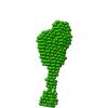


 Search similar-shape structures of this assembly by Omokage search (details)
 DORIS III X33


