 Movie
Movie Controller
 Structure viewers
 About Yorodumi Papers


+Search query
-Structure paper
| Title | Characterization of a fluorophore binding RNA aptamer by fluorescence correlation spectroscopy and small angle X-ray scattering. |
|---|---|
| Journal, issue, pages | Anal Biochem, Vol. 389, Issue 1, Page 52-62, Year 2009 |
| Publish date | Jun 1, 2009 |
 Authors Authors | Arne Werner / Petr V Konarev / Dmitri I Svergun / Ulrich Hahn /  |
| PubMed Abstract | Using fluorescence correlation spectroscopy (FCS), we have established an in vitro assay to study RNA dynamics by analyzing fluorophore binding RNA aptamers at the single molecule level. The RNA ...Using fluorescence correlation spectroscopy (FCS), we have established an in vitro assay to study RNA dynamics by analyzing fluorophore binding RNA aptamers at the single molecule level. The RNA aptamer SRB2m, a minimized variant of the initially selected aptamer SRB-2, has a high affinity to the disulfonated triphenylmethane dye sulforhodamine B. A mobility shift of sulforhodamine B after binding to SRB2m was measured. In contrast, patent blue V (PBV) is visible only if complexed with SRB2m due to increased molecular brightness and minimal background. With small angle X-ray scattering (SAXS), the three-dimensional structure of the RNA aptamer was characterized at low resolution to analyze the effect of fluorophore binding. The aptamer and sulforhodamine B-aptamer complex was found to be predominantly dimeric in solution. Interaction of PBV with SRB2m led to a dissociation of SRB2m dimers into monomers. Radii of gyration and hydrodynamic radii, gained from dynamic light scattering, FCS, and fluorescence cross-correlation experiments, led to comparable conclusions. Our study demonstrates how RNA-aptamer fluorophore complexes can be simultaneously structurally and photophysically characterized by FCS. Furthermore, fluorophore binding RNA aptamers provide a tool for visualizing single RNA molecules. |
 External links External links | Anal Biochem / PubMed:19303859 |
| Methods | SAS (X-ray synchrotron) |
| Structure data | 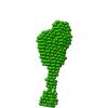 SASDA54:  SASDA74: 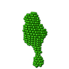 SASDA84: |
