 Movie
Movie Controller


+ Open data

- Basic information
| Entry | Database: PDB / ID: 3sok | ||||||
|---|---|---|---|---|---|---|---|
| Title | Dichelobacter nodosus pilin FimA | ||||||
 Components Components | Fimbrial protein | ||||||
 Keywords Keywords | CELL ADHESION / pilus subunit / extracellular | ||||||
| Function / homology |  Function and homology information Function and homology information | ||||||
| Biological species |  Dichelobacter nodosus (bacteria) Dichelobacter nodosus (bacteria) | ||||||
| Method |  X-RAY DIFFRACTION / SYNCHROTRON / MOLECULAR REPLACEMENT / Resolution: 2.3 Å X-RAY DIFFRACTION / SYNCHROTRON / MOLECULAR REPLACEMENT / Resolution: 2.3 Å | ||||||
 Authors Authors | Arvai, A.S. / Craig, L. / Hartung, S. / Wood, T. / Kolappan, S. / Shin, D.S. / Tainer, J.A. | ||||||
 Citation Citation | Journal: J.Biol.Chem. / Year: 2011 Title: Ultrahigh Resolution and Full-length Pilin Structures with Insights for Filament Assembly, Pathogenic Functions, and Vaccine Potential. Authors: Hartung, S. / Arvai, A.S. / Wood, T. / Kolappan, S. / Shin, D.S. / Craig, L. / Tainer, J.A. | ||||||
| History |
|
- Structure visualization
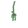
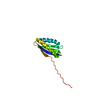
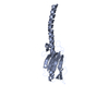
| Structure viewer | Molecule: MolmilJmol/JSmol |
|---|
- Downloads & links
-Download
| PDBx/mmCIF format | 3sok.cif.gz | 121 KB | Display | PDBx/mmCIF format |
|---|---|---|---|---|
| PDB format | pdb3sok.ent.gz | 95.7 KB | Display | PDB format |
| PDBx/mmJSON format | 3sok.json.gz | Tree view | PDBx/mmJSON format | |
| Others |  Other downloads Other downloads |
-Validation report
| Arichive directory | https://data.pdbj.org/pub/pdb/validation_reports/so/3sokftp://data.pdbj.org/pub/pdb/validation_reports/so/3sok | HTTPS FTP |
|---|
-Related structure data
| Related structure data |  3sojC 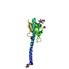 2hi2S C: citing same article ( S: Starting model for refinement |
|---|---|
| Similar structure data |


-Links
 PDBj
PDBj
- Assembly
| Deposited unit | 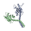
| ||||||||
|---|---|---|---|---|---|---|---|---|---|
| 1 | 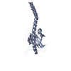
| ||||||||
| 2 | 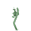
| ||||||||
| Unit cell |
|
-Components
| #1: Protein | Mass: 16108.239 Da / Num. of mol.: 2 Source method: isolated from a genetically manipulated source Source: (gene. exp.) Dichelobacter nodosus (bacteria) / Gene: fimA / Plasmid: pJSM202 / Production host:  Pseudomonas aeruginosa (bacteria) / Strain (production host): PAK / References: UniProt: P02975 Pseudomonas aeruginosa (bacteria) / Strain (production host): PAK / References: UniProt: P02975#2: Water | ChemComp-HOH / |  Mass: 18.015 Da / Num. of mol.: 61 / Source method: isolated from a natural source / Formula: H2O Mass: 18.015 Da / Num. of mol.: 61 / Source method: isolated from a natural source / Formula: H2OHas protein modification | Y | |
|---|
-Experimental details
-Experiment
| Experiment | Method: X-RAY DIFFRACTION / Number of used crystals: 1 |
|---|
- Sample preparation
| Crystal | Density Matthews: 3.2 Å3/Da / Density % sol: 61.6 % |
|---|---|
| Crystal grow | Temperature: 295 K / Method: vapor diffusion, hanging drop / pH: 6.6 Details: 60% saturated ammonium sulfate 5% saturated NaCl 100mM Na Citrate 5% 1M NaPO4, pH 9.23, VAPOR DIFFUSION, HANGING DROP, temperature 295K |
-Data collection
| Diffraction | Mean temperature: 100 K |
|---|---|
| Diffraction source | Source: SYNCHROTRON / Site: SSRL  / Beamline: BL9-1 / Wavelength: 1.1 Å / Beamline: BL9-1 / Wavelength: 1.1 Å |
| Detector | Type: ADSC QUANTUM 315r / Detector: CCD / Date: Mar 14, 2008 |
| Radiation | Monochromator: Side-scattering cuberoot I-beam bent single crystal Protocol: SINGLE WAVELENGTH / Monochromatic (M) / Laue (L): M / Scattering type: x-ray |
| Radiation wavelength | Wavelength: 1.1 Å / Relative weight: 1 |
| Reflection | Resolution: 2→47.3 Å / Num. all: 39396 / Num. obs: 27380 / % possible obs: 96.5 % / Observed criterion σ(F): 1 / Observed criterion σ(I): 1 |
| Reflection shell | Resolution: 2.3→2.52 Å / % possible all: 97.1 |
- Processing
| Software |
| ||||||||||||||||||||||||||||||||||||||||||||||||||||||||||||||||||||||||||||||||||||||||||||||||||
|---|---|---|---|---|---|---|---|---|---|---|---|---|---|---|---|---|---|---|---|---|---|---|---|---|---|---|---|---|---|---|---|---|---|---|---|---|---|---|---|---|---|---|---|---|---|---|---|---|---|---|---|---|---|---|---|---|---|---|---|---|---|---|---|---|---|---|---|---|---|---|---|---|---|---|---|---|---|---|---|---|---|---|---|---|---|---|---|---|---|---|---|---|---|---|---|---|---|---|---|
| Refinement | Method to determine structure: MOLECULAR REPLACEMENT Starting model: PDB code 2HI2 conserved core (residues 1-55 + 78-92 + 105-111) Resolution: 2.3→47.283 Å / SU ML: 0.85 / σ(F): 1 / Phase error: 28.78 / Stereochemistry target values: ML
| ||||||||||||||||||||||||||||||||||||||||||||||||||||||||||||||||||||||||||||||||||||||||||||||||||
| Solvent computation | Shrinkage radii: 0.72 Å / VDW probe radii: 1 Å / Solvent model: FLAT BULK SOLVENT MODEL / Bsol: 49.362 Å2 / ksol: 0.352 e/Å3 | ||||||||||||||||||||||||||||||||||||||||||||||||||||||||||||||||||||||||||||||||||||||||||||||||||
| Displacement parameters |
| ||||||||||||||||||||||||||||||||||||||||||||||||||||||||||||||||||||||||||||||||||||||||||||||||||
| Refinement step | Cycle: LAST / Resolution: 2.3→47.283 Å
| ||||||||||||||||||||||||||||||||||||||||||||||||||||||||||||||||||||||||||||||||||||||||||||||||||
| Refine LS restraints |
| ||||||||||||||||||||||||||||||||||||||||||||||||||||||||||||||||||||||||||||||||||||||||||||||||||
| LS refinement shell |
| ||||||||||||||||||||||||||||||||||||||||||||||||||||||||||||||||||||||||||||||||||||||||||||||||||
| Refinement TLS params. | Method: refined / Origin x: -21.7093 Å / Origin y: 0.0057 Å / Origin z: 12.6855 Å
| ||||||||||||||||||||||||||||||||||||||||||||||||||||||||||||||||||||||||||||||||||||||||||||||||||
| Refinement TLS group | Selection details: all |
