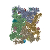
Movie
Movie Controller


+ Open data

- Basic information
| Entry | Database: PDB / ID: 9s5e | |||||||||||||||||||||||||||
|---|---|---|---|---|---|---|---|---|---|---|---|---|---|---|---|---|---|---|---|---|---|---|---|---|---|---|---|---|
| Title | Tight class, Apo-state RyR1 in the native membrane solved by StA | |||||||||||||||||||||||||||
 Components Components |
| |||||||||||||||||||||||||||
 Keywords Keywords | MEMBRANE PROTEIN / ryanodine receptor type 1 / ryr1 / skeletal muscle / sarcoplasmic reticulum / SR / native membrane | |||||||||||||||||||||||||||
| Function / homology |  Function and homology information Function and homology informationATP-gated ion channel activity / cytoplasmic side of membrane / ryanodine-sensitive calcium-release channel activity / terminal cisterna / release of sequestered calcium ion into cytosol by sarcoplasmic reticulum / ossification involved in bone maturation / cellular response to caffeine / skin development / ryanodine receptor complex / organelle membrane ...ATP-gated ion channel activity / cytoplasmic side of membrane / ryanodine-sensitive calcium-release channel activity / terminal cisterna / release of sequestered calcium ion into cytosol by sarcoplasmic reticulum / ossification involved in bone maturation / cellular response to caffeine / skin development / ryanodine receptor complex / organelle membrane / outflow tract morphogenesis / smooth endoplasmic reticulum / intracellularly gated calcium channel activity / intracellular membrane-bounded organelle / regulation of ryanodine-sensitive calcium-release channel activity / toxic substance binding / skeletal muscle fiber development / voltage-gated calcium channel activity / release of sequestered calcium ion into cytosol / regulation of release of sequestered calcium ion into cytosol by sarcoplasmic reticulum / cellular response to calcium ion / muscle contraction / sarcoplasmic reticulum membrane / peptidylprolyl isomerase / sarcoplasmic reticulum / peptidyl-prolyl cis-trans isomerase activity / striated muscle contraction / sarcolemma / intracellular calcium ion homeostasis / calcium channel activity / Z disc / calcium ion transmembrane transport / disordered domain specific binding / calmodulin binding / protein homotetramerization / transmembrane transporter binding / calcium ion binding / ATP binding / membrane / identical protein binding / cytosol Similarity search - Function | |||||||||||||||||||||||||||
| Biological species |   Oryctolagus cuniculus (rabbit) Oryctolagus cuniculus (rabbit) | |||||||||||||||||||||||||||
| Method | ELECTRON MICROSCOPY / subtomogram averaging / cryo EM / Resolution: 6.99 Å | |||||||||||||||||||||||||||
 Authors Authors | Mikirtumov, V. | |||||||||||||||||||||||||||
| Funding support |  Germany, 2items Germany, 2items
| |||||||||||||||||||||||||||
 Citation Citation | Journal: Nat Commun / Year: 2026 Title: Ligand-induced activation of RyR1 in native membranes. Authors: Vasilii Mikirtumov / Sabrina Golusik / Ruifeng Huo / Thiemo Sprink / Nikita Balyschew / Wen Yang / Christoph Diebolder / Shuguang Yuan / Abhay Kotecha / Mikhail Kudryashev /    Abstract: Synchronized calcium release through arrays of the ryanodine receptor RyR1, fundamental to skeletal muscle excitation-contraction coupling, is achieved through the mechanical interaction of RyR1s and ...Synchronized calcium release through arrays of the ryanodine receptor RyR1, fundamental to skeletal muscle excitation-contraction coupling, is achieved through the mechanical interaction of RyR1s and voltage-sensing receptors DHPR that activate RyR1s in response to action potentials. The calcium release is enhanced through "coupled gating", when the activation of one channel promotes the opening of its neighbours. Here, we determine high-resolution structures of RyR1 in native sarcoplasmic reticulum membranes by cryo-EM/ET, capturing the conformations along the activation pathway and corner-to-corner interfaces between adjacent RyR1 receptors. Compared with purified RyR1s, receptors in native membranes follow an activation pathway with reduced cytosolic-shell tilt and greater consecutive in-plane rotation. Activation-induced rotation remodels the inter-receptor interface, lowering the energy barrier to the cooperative opening of the receptor cluster. Our analysis demonstrates how the native membrane receptor lattice influences ion channel cluster dynamics and provides a mechanistic framework for understanding calcium signaling in muscle. | |||||||||||||||||||||||||||
| History |
|
- Structure visualization
| Structure viewer | Molecule: MolmilJmol/JSmol |
|---|
- Downloads & links
-Download
| PDBx/mmCIF format | 9s5e.cif.gz | 5.7 MB | Display | PDBx/mmCIF format |
|---|---|---|---|---|
| PDB format | pdb9s5e.ent.gz | Display | PDB format | |
| PDBx/mmJSON format | 9s5e.json.gz | Tree view | PDBx/mmJSON format | |
| Others |  Other downloads Other downloads |
-Validation report
| Arichive directory | https://data.pdbj.org/pub/pdb/validation_reports/s5/9s5eftp://data.pdbj.org/pub/pdb/validation_reports/s5/9s5e | HTTPS FTP |
|---|
-Related structure data
| Related structure data |  54595MC  9s16C  9s1hC  9s2rC  9s2sC  9s3uC  9s3vC  9s4lC  9s4nC  9s54C  9s55C  9s5aC  9s5cC  9s5fC  9s5wC  9s5yC  9s5zC M: map data used to model this data C: citing same article ( |
|---|---|
| Similar structure data |


-Links
 PDBj
PDBj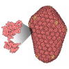

- Assembly
| Deposited unit | 
|
|---|---|
| 1 |
|
-Components
| #1: Protein | Mass: 565908.625 Da / Num. of mol.: 4 / Source method: isolated from a natural source / Source: (natural) Oryctolagus cuniculus (rabbit) / References: UniProt: P11716#2: Protein | Mass: 11967.705 Da / Num. of mol.: 4 / Source method: isolated from a natural source / Source: (natural) Oryctolagus cuniculus (rabbit) / References: UniProt: P62943, peptidylprolyl isomeraseHas protein modification | Y | |
|---|
-Experimental details
-Experiment
| Experiment | Method: ELECTRON MICROSCOPY |
|---|---|
| EM experiment | Aggregation state: PARTICLE / 3D reconstruction method: subtomogram averaging |
- Sample preparation
| Component | Name: Ryanodine receptor type 1 in complex with FKBP12 in the sarcoplasmic reticulum membrane Type: ORGANELLE OR CELLULAR COMPONENT Details: Sarcoplasmic reticulum membrane vesicles purified from the native skeletal muscle tissue Entity ID: all / Source: NATURAL |
|---|---|
| Molecular weight | Value: 2.2 MDa / Experimental value: NO |
| Source (natural) | Organism: Oryctolagus cuniculus (rabbit) / Organ: hind leg and back / Organelle: sarcoplasmic reticulum / Tissue: skeletal muscle |
| Buffer solution | pH: 7.1 |
| Specimen | Conc.: 29.5 mg/ml / Embedding applied: NO / Shadowing applied: NO / Staining applied: NO / Vitrification applied: YES |
| Vitrification | Instrument: FEI VITROBOT MARK IV / Cryogen name: ETHANE / Humidity: 95 % / Chamber temperature: 277.15 K |
- Electron microscopy imaging
| Experimental equipment |  Model: Titan Krios / Image courtesy: FEI Company |
|---|---|
| Microscopy | Model: TFS KRIOS |
| Electron gun | Electron source:  FIELD EMISSION GUN / Accelerating voltage: 300 kV / Illumination mode: FLOOD BEAM FIELD EMISSION GUN / Accelerating voltage: 300 kV / Illumination mode: FLOOD BEAM |
| Electron lens | Mode: BRIGHT FIELD / Nominal magnification: 64000 X / Calibrated magnification: 64000 X / Nominal defocus max: 5000 nm / Nominal defocus min: 3000 nm / Calibrated defocus min: 1800 nm / Calibrated defocus max: 3800 nm / Cs: 2.7 mm / C2 aperture diameter: 50 µm |
| Specimen holder | Cryogen: NITROGEN / Specimen holder model: FEI TITAN KRIOS AUTOGRID HOLDER |
| Image recording | Average exposure time: 1.88 sec. / Electron dose: 4 e/Å2 / Avg electron dose per subtomogram: 132 e/Å2 / Film or detector model: TFS FALCON 4i (4k x 4k) / Num. of real images: 1 |
| EM imaging optics | Energyfilter name: TFS Selectris X / Energyfilter slit width: 10 eV |
| Image scans | Width: 8192 / Height: 8192 |
- Processing
| EM software |
| ||||||||||||||||||||||||||||||||||||||||||||||||||||
|---|---|---|---|---|---|---|---|---|---|---|---|---|---|---|---|---|---|---|---|---|---|---|---|---|---|---|---|---|---|---|---|---|---|---|---|---|---|---|---|---|---|---|---|---|---|---|---|---|---|---|---|---|---|
| CTF correction | Type: PHASE FLIPPING AND AMPLITUDE CORRECTION | ||||||||||||||||||||||||||||||||||||||||||||||||||||
| Particle selection | Num. of particles selected: 2171564 | ||||||||||||||||||||||||||||||||||||||||||||||||||||
| Symmetry | Point symmetry: C4 (4 fold cyclic) | ||||||||||||||||||||||||||||||||||||||||||||||||||||
| 3D reconstruction | Resolution: 6.99 Å / Resolution method: FSC 0.143 CUT-OFF / Num. of particles: 1088 / Algorithm: FOURIER SPACE / Num. of class averages: 1 / Symmetry type: POINT | ||||||||||||||||||||||||||||||||||||||||||||||||||||
| EM volume selection | Method: Dynamo-GPU TomoBEAR Template Matching / Num. of tomograms: 194 / Num. of volumes extracted: 44333 / Reference model: EMD-17272 | ||||||||||||||||||||||||||||||||||||||||||||||||||||
| Atomic model building | Protocol: FLEXIBLE FIT / Space: REAL | ||||||||||||||||||||||||||||||||||||||||||||||||||||
| Atomic model building | PDB-ID: 7tzc Accession code: 7tzc / Source name: PDB / Type: experimental model |