Movie
Movie Controller


+ Open data

- Basic information
| Entry | Database: PDB / ID: 9s16 | |||||||||||||||
|---|---|---|---|---|---|---|---|---|---|---|---|---|---|---|---|---|
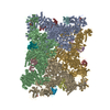
| Title | Apo-state RyR1 in the native membrane solved by "SPA | |||||||||||||||
 Components Components |
| |||||||||||||||
 Keywords Keywords | MEMBRANE PROTEIN / ryanodine receptor type 1 / ryr1 / skeletal muscle / sarcoplasmic reticulum / SR / native membrane | |||||||||||||||
| Function / homology |  Function and homology information Function and homology informationATP-gated ion channel activity / cytoplasmic side of membrane / ryanodine-sensitive calcium-release channel activity / terminal cisterna / release of sequestered calcium ion into cytosol by sarcoplasmic reticulum / ossification involved in bone maturation / cellular response to caffeine / skin development / ryanodine receptor complex / organelle membrane ...ATP-gated ion channel activity / cytoplasmic side of membrane / ryanodine-sensitive calcium-release channel activity / terminal cisterna / release of sequestered calcium ion into cytosol by sarcoplasmic reticulum / ossification involved in bone maturation / cellular response to caffeine / skin development / ryanodine receptor complex / organelle membrane / outflow tract morphogenesis / smooth endoplasmic reticulum / intracellularly gated calcium channel activity / intracellular membrane-bounded organelle / regulation of ryanodine-sensitive calcium-release channel activity / toxic substance binding / skeletal muscle fiber development / voltage-gated calcium channel activity / release of sequestered calcium ion into cytosol / regulation of release of sequestered calcium ion into cytosol by sarcoplasmic reticulum / cellular response to calcium ion / muscle contraction / sarcoplasmic reticulum membrane / peptidylprolyl isomerase / sarcoplasmic reticulum / peptidyl-prolyl cis-trans isomerase activity / striated muscle contraction / sarcolemma / intracellular calcium ion homeostasis / calcium channel activity / Z disc / calcium ion transmembrane transport / disordered domain specific binding / calmodulin binding / protein homotetramerization / transmembrane transporter binding / calcium ion binding / ATP binding / membrane / identical protein binding / cytosol Similarity search - Function | |||||||||||||||
| Biological species |   Oryctolagus cuniculus (rabbit) Oryctolagus cuniculus (rabbit) | |||||||||||||||
| Method | ELECTRON MICROSCOPY / single particle reconstruction / cryo EM / Resolution: 3.6 Å | |||||||||||||||
 Authors Authors | Mikirtumov, V. | |||||||||||||||
| Funding support |  Germany, 2items Germany, 2items
| |||||||||||||||
 Citation Citation | Journal: To Be Published Title: Activation of RyR1 in native membranes Authors: Mikirtumov, V. / Golusik, S. / Sprink, T. / Balyschew, N. / Yang, W. / Kotecha, A. / Kudryashev, M. | |||||||||||||||
| History |
|
- Structure visualization
| Structure viewer | Molecule: MolmilJmol/JSmol |
|---|
- Downloads & links
-Download
| PDBx/mmCIF format | 9s16.cif.gz | 5.7 MB | Display | PDBx/mmCIF format |
|---|---|---|---|---|
| PDB format | pdb9s16.ent.gz | Display | PDB format | |
| PDBx/mmJSON format | 9s16.json.gz | Tree view | PDBx/mmJSON format | |
| Others |  Other downloads Other downloads |
-Validation report
| Arichive directory | https://data.pdbj.org/pub/pdb/validation_reports/s1/9s16ftp://data.pdbj.org/pub/pdb/validation_reports/s1/9s16 | HTTPS FTP |
|---|
-Related structure data
| Related structure data |  54445MC  9s1hC  9s2rC  9s2sC  9s3uC  9s3vC  9s4lC  9s4nC  9s54C  9s55C  9s5aC  9s5cC  9s5eC  9s5fC  9s5wC  9s5yC  9s5zC C: citing same article ( M: map data used to model this data |
|---|---|
| Similar structure data |


-Links
 PDBj
PDBj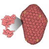

- Assembly
| Deposited unit | 
|
|---|---|
| 1 |
|
-Components
| #1: Protein | Mass: 565908.625 Da / Num. of mol.: 4 / Source method: isolated from a natural source / Source: (natural) Oryctolagus cuniculus (rabbit) / Tissue: skeletal muscle from back and hind leg / References: UniProt: P11716#2: Protein | Mass: 11967.705 Da / Num. of mol.: 4 / Source method: isolated from a natural source / Source: (natural) Oryctolagus cuniculus (rabbit) / References: UniProt: P62943, peptidylprolyl isomerase#3: Chemical | ChemComp-POV / ( 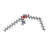 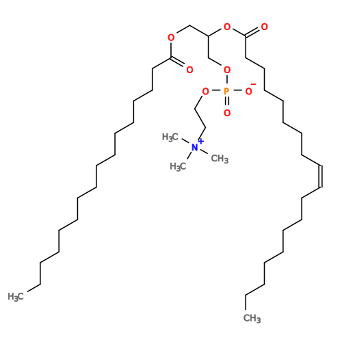 Mass: 760.076 Da / Num. of mol.: 8 / Source method: obtained synthetically / Formula: C42H82NO8P / Feature type: SUBJECT OF INVESTIGATION / Comment: phospholipid*YM Mass: 760.076 Da / Num. of mol.: 8 / Source method: obtained synthetically / Formula: C42H82NO8P / Feature type: SUBJECT OF INVESTIGATION / Comment: phospholipid*YM#4: Chemical | ChemComp-ZN /   Mass: 65.409 Da / Num. of mol.: 4 / Source method: obtained synthetically / Formula: Zn / Feature type: SUBJECT OF INVESTIGATION Mass: 65.409 Da / Num. of mol.: 4 / Source method: obtained synthetically / Formula: Zn / Feature type: SUBJECT OF INVESTIGATIONHas ligand of interest | Y | Has protein modification | Y | |
|---|
-Experimental details
-Experiment
| Experiment | Method: ELECTRON MICROSCOPY |
|---|---|
| EM experiment | Aggregation state: PARTICLE / 3D reconstruction method: single particle reconstruction |
- Sample preparation
| Component | Name: Ryanodine receptor type 1 in complex with FKBP12 in the sarcoplasmic reticulum membrane Type: ORGANELLE OR CELLULAR COMPONENT Details: Sarcoplasmic reticulum membrane vesicles purified from the native skeletal muscle tissue Entity ID: #1-#2 / Source: NATURAL |
|---|---|
| Molecular weight | Value: 2.2 MDa / Experimental value: NO |
| Source (natural) | Organism: Oryctolagus cuniculus (rabbit) / Organ: hind leg and back / Organelle: sarcoplasmic reticulum / Tissue: skeletal muscle |
| Buffer solution | pH: 7.1 |
| Specimen | Conc.: 29.5 mg/ml / Embedding applied: NO / Shadowing applied: NO / Staining applied: NO / Vitrification applied: YES |
| Specimen support | Grid material: GOLD / Grid mesh size: 200 divisions/in. / Grid type: UltrAuFoil R2/2 |
| Vitrification | Instrument: FEI VITROBOT MARK IV / Cryogen name: ETHANE / Humidity: 95 % / Chamber temperature: 277.15 K |
- Electron microscopy imaging
| Experimental equipment |  Model: Titan Krios / Image courtesy: FEI Company |
|---|---|
| Microscopy | Model: TFS KRIOS |
| Electron gun | Electron source:  FIELD EMISSION GUN / Accelerating voltage: 300 kV / Illumination mode: FLOOD BEAM FIELD EMISSION GUN / Accelerating voltage: 300 kV / Illumination mode: FLOOD BEAM |
| Electron lens | Mode: BRIGHT FIELD / Nominal magnification: 64000 X / Calibrated magnification: 64000 X / Nominal defocus max: 4000 nm / Nominal defocus min: 500 nm / Calibrated defocus min: 500 nm / Calibrated defocus max: 4000 nm / Cs: 2.7 mm / C2 aperture diameter: 50 µm |
| Specimen holder | Cryogen: NITROGEN / Specimen holder model: FEI TITAN KRIOS AUTOGRID HOLDER |
| Image recording | Average exposure time: 1.8 sec. / Electron dose: 60.9 e/Å2 / Film or detector model: GATAN K3 (6k x 4k) / Num. of grids imaged: 1 / Num. of real images: 6204 |
| EM imaging optics | Energyfilter name: GIF Bioquantum / Energyfilter slit width: 20 eV |
| Image scans | Width: 11520 / Height: 8184 |
- Processing
| EM software |
| ||||||||||||||||||||||||||||||||||||||||
|---|---|---|---|---|---|---|---|---|---|---|---|---|---|---|---|---|---|---|---|---|---|---|---|---|---|---|---|---|---|---|---|---|---|---|---|---|---|---|---|---|---|
| CTF correction | Type: PHASE FLIPPING AND AMPLITUDE CORRECTION | ||||||||||||||||||||||||||||||||||||||||
| Particle selection | Num. of particles selected: 1480995 | ||||||||||||||||||||||||||||||||||||||||
| Symmetry | Point symmetry: C4 (4 fold cyclic) | ||||||||||||||||||||||||||||||||||||||||
| 3D reconstruction | Resolution: 3.6 Å / Resolution method: FSC 0.143 CUT-OFF / Num. of particles: 25786 / Algorithm: FOURIER SPACE Details: The composite map consists of the four part with resolutions 3.7, 4.7, 3.9 and 3.6 Angstrom Num. of class averages: 1 / Symmetry type: POINT | ||||||||||||||||||||||||||||||||||||||||
| Atomic model building | Protocol: FLEXIBLE FIT / Space: REAL | ||||||||||||||||||||||||||||||||||||||||
| Atomic model building | PDB-ID: 7tzc Accession code: 7tzc / Source name: PDB / Type: experimental model |