ムービー
ムービー コントローラー


+ データを開く

- 基本情報
基本情報
| 登録情報 |  | |||||||||
|---|---|---|---|---|---|---|---|---|---|---|
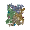
| タイトル | Focused map of the CSol activation core part, Apo-state RyR1 in the native membrane solved by "SPA" | |||||||||
 マップデータ マップデータ | ||||||||||
 試料 試料 |
| |||||||||
 キーワード キーワード | ryanodine receptor type 1 / ryr1 / skeletal muscle / sarcoplasmic reticulum / SR / native membrane / MEMBRANE PROTEIN | |||||||||
| 生物種 |   Oryctolagus cuniculus (ウサギ) Oryctolagus cuniculus (ウサギ) | |||||||||
| 手法 | 単粒子再構成法 / クライオ電子顕微鏡法 / 解像度: 3.93 Å | |||||||||
 データ登録者 データ登録者 | Mikirtumov V | |||||||||
| 資金援助 |  ドイツ, 2件 ドイツ, 2件
| |||||||||
 引用 引用 | ジャーナル: Nat Commun / 年: 2026 タイトル: Ligand-induced activation of RyR1 in native membranes. 著者: Vasilii Mikirtumov / Sabrina Golusik / Ruifeng Huo / Thiemo Sprink / Nikita Balyschew / Wen Yang / Christoph Diebolder / Shuguang Yuan / Abhay Kotecha / Mikhail Kudryashev /    要旨: Synchronized calcium release through arrays of the ryanodine receptor RyR1, fundamental to skeletal muscle excitation-contraction coupling, is achieved through the mechanical interaction of RyR1s and ...Synchronized calcium release through arrays of the ryanodine receptor RyR1, fundamental to skeletal muscle excitation-contraction coupling, is achieved through the mechanical interaction of RyR1s and voltage-sensing receptors DHPR that activate RyR1s in response to action potentials. The calcium release is enhanced through "coupled gating", when the activation of one channel promotes the opening of its neighbours. Here, we determine high-resolution structures of RyR1 in native sarcoplasmic reticulum membranes by cryo-EM/ET, capturing the conformations along the activation pathway and corner-to-corner interfaces between adjacent RyR1 receptors. Compared with purified RyR1s, receptors in native membranes follow an activation pathway with reduced cytosolic-shell tilt and greater consecutive in-plane rotation. Activation-induced rotation remodels the inter-receptor interface, lowering the energy barrier to the cooperative opening of the receptor cluster. Our analysis demonstrates how the native membrane receptor lattice influences ion channel cluster dynamics and provides a mechanistic framework for understanding calcium signaling in muscle. | |||||||||
| 履歴 |
|
- 構造の表示
構造の表示
| 添付画像 |
|---|

- ダウンロードとリンク
-EMDBアーカイブ
| マップデータ | emd_54435.map.gz | 306.4 MB |  EMDBマップデータ形式 EMDBマップデータ形式 | |
|---|---|---|---|---|
| ヘッダ (付随情報) | emd-54435-v30.xmlemd-54435.xml | 19.7 KB 19.7 KB | 表示 表示 | EMDBヘッダ |
| 画像 |  emd_54435.png emd_54435.png | 64.8 KB | ||
| マスクデータ | emd_54435_msk_1.map | 325 MB | マスクマップ | |
| Filedesc metadata | emd-54435.cif.gz | 5.2 KB | ||
| その他 | emd_54435_half_map_1.map.gzemd_54435_half_map_2.map.gz | 301.3 MB 301.3 MB | ||
| アーカイブディレクトリ |  https://data.pdbj.org/pub/emdb/structures/EMD-54435ftp://data.pdbj.org/pub/emdb/structures/EMD-54435 https://data.pdbj.org/pub/emdb/structures/EMD-54435ftp://data.pdbj.org/pub/emdb/structures/EMD-54435 | HTTPS FTP |
-関連構造データ
| 関連構造データ |  9s16C  9s1hC  9s2rC  9s2sC  9s3uC  9s3vC  9s4lC  9s4nC  9s54C  9s55C  9s5aC  9s5cC  9s5eC  9s5fC  9s5wC  9s5yC  9s5zC C: 同じ文献を引用 ( |
|---|


-リンク
| EMDBのページ | EMDB (EBI/PDBe) / EMDataResource |
|---|
-マップ
| ファイル | ダウンロード / ファイル: emd_54435.map.gz / 形式: CCP4 / 大きさ: 325 MB / タイプ: IMAGE STORED AS FLOATING POINT NUMBER (4 BYTES) | ||||||||||||||||||||||||||||||||||||
|---|---|---|---|---|---|---|---|---|---|---|---|---|---|---|---|---|---|---|---|---|---|---|---|---|---|---|---|---|---|---|---|---|---|---|---|---|---|
| 投影像・断面図 | 画像のコントロール
画像は Spider により作成 | ||||||||||||||||||||||||||||||||||||
| ボクセルのサイズ | X=Y=Z: 1.38 Å | ||||||||||||||||||||||||||||||||||||
| 密度 |
| ||||||||||||||||||||||||||||||||||||
| 対称性 | 空間群: 1 | ||||||||||||||||||||||||||||||||||||
| 詳細 | EMDB XML:
|
 Z (Sec.)
 Y (Row.)
 X (Col.)


-添付データ
-マスク #1
| ファイル | emd_54435_msk_1.map | ||||||||||||
|---|---|---|---|---|---|---|---|---|---|---|---|---|---|
| 投影像・断面図 |
| ||||||||||||
| 密度ヒストグラム |


-ハーフマップ: #1
| ファイル | emd_54435_half_map_1.map | ||||||||||||
|---|---|---|---|---|---|---|---|---|---|---|---|---|---|
| 投影像・断面図 |
| ||||||||||||
| 密度ヒストグラム |


-ハーフマップ: #2
| ファイル | emd_54435_half_map_2.map | ||||||||||||
|---|---|---|---|---|---|---|---|---|---|---|---|---|---|
| 投影像・断面図 |
| ||||||||||||
| 密度ヒストグラム |


- 試料の構成要素
-全体 : Ryanodine receptor type 1 in complex with FKBP12 in the sarcoplas...
| 全体 | 名称: Ryanodine receptor type 1 in complex with FKBP12 in the sarcoplasmic reticulum membrane |
|---|---|
| 要素 |
|
-超分子 #1: Ryanodine receptor type 1 in complex with FKBP12 in the sarcoplas...
| 超分子 | 名称: Ryanodine receptor type 1 in complex with FKBP12 in the sarcoplasmic reticulum membrane タイプ: organelle_or_cellular_component / ID: 1 / 親要素: 0 / 含まれる分子: #1-#2 詳細: Sarcoplasmic reticulum membrane vesicles purified from the native skeletal muscle tissue |
|---|---|
| 由来(天然) | 生物種: Oryctolagus cuniculus (ウサギ) / 器官: hind leg and back / 組織: skeletal muscle / Organelle: sarcoplasmic reticulum |
-実験情報
-構造解析
| 手法 | クライオ電子顕微鏡法 |
|---|---|
 解析 解析 | 単粒子再構成法 |
| 試料の集合状態 | particle |
-試料調製
| 濃度 | 29.5 mg/mL |
|---|---|
| 緩衝液 | pH: 7.1 |
| グリッド | モデル: UltrAuFoil R2/2 / 材質: GOLD / メッシュ: 200 / 支持フィルム - 材質: GOLD / 支持フィルム - トポロジー: HOLEY ARRAY |
| 凍結 | 凍結剤: ETHANE / チャンバー内湿度: 95 % / チャンバー内温度: 277.15 K / 装置: FEI VITROBOT MARK IV |
- 電子顕微鏡法
| 顕微鏡 | TFS KRIOS |
|---|---|
| 特殊光学系 | エネルギーフィルター - 名称: GIF Bioquantum / エネルギーフィルター - スリット幅: 20 eV |
| 撮影 | フィルム・検出器のモデル: GATAN K3 (6k x 4k) / デジタル化 - サイズ - 横: 11520 pixel / デジタル化 - サイズ - 縦: 8184 pixel / 撮影したグリッド数: 1 / 実像数: 6204 / 平均露光時間: 1.8 sec. / 平均電子線量: 60.9 e/Å2 |
| 電子線 | 加速電圧: 300 kV / 電子線源:  FIELD EMISSION GUN FIELD EMISSION GUN |
| 電子光学系 | C2レンズ絞り径: 50.0 µm / 最大 デフォーカス(補正後): 4.0 µm / 最小 デフォーカス(補正後): 0.5 µm / 倍率(補正後): 64000 / 照射モード: FLOOD BEAM / 撮影モード: BRIGHT FIELD / Cs: 2.7 mm / 最大 デフォーカス(公称値): 4.0 µm / 最小 デフォーカス(公称値): 0.5 µm / 倍率(公称値): 64000 |
| 試料ステージ | 試料ホルダーモデル: FEI TITAN KRIOS AUTOGRID HOLDER ホルダー冷却材: NITROGEN |
| 実験機器 |  モデル: Titan Krios / 画像提供: FEI Company |
+画像解析
-原子モデル構築 1
| 初期モデル | PDB ID: Chain - Source name: PDB / Chain - Initial model type: experimental model |
|---|---|
| 精密化 | 空間: REAL / プロトコル: FLEXIBLE FIT |