 Movie
Movie Controller

[English] 日本語
 Yorodumi
- PDB-8vrj: Rigid body fitted model for gamma tubulin ring complex capped mic... -
+ Open data

- Basic information
| Entry | Database: PDB / ID: 8vrj | ||||||
|---|---|---|---|---|---|---|---|
| Title | Rigid body fitted model for gamma tubulin ring complex capped microtubule | ||||||
 Components Components |
| ||||||
 Keywords Keywords | CELL CYCLE / Microtubule nucleation complex bound to a microtubule | ||||||
| Function / homology |  Function and homology information Function and homology informationmicrotubule nucleation by interphase microtubule organizing center / netrin receptor binding / gamma-tubulin complex localization / microtubule nucleator activity / dorsal root ganglion development / Post-chaperonin tubulin folding pathway / Cilium Assembly / positive regulation of norepinephrine uptake / cytoskeleton-dependent intracellular transport / polar microtubule ...microtubule nucleation by interphase microtubule organizing center / netrin receptor binding / gamma-tubulin complex localization / microtubule nucleator activity / dorsal root ganglion development / Post-chaperonin tubulin folding pathway / Cilium Assembly / positive regulation of norepinephrine uptake / cytoskeleton-dependent intracellular transport / polar microtubule / Carboxyterminal post-translational modifications of tubulin / Microtubule-dependent trafficking of connexons from Golgi to the plasma membrane / interphase microtubule organizing center / gamma-tubulin complex / gamma-tubulin ring complex / mitotic spindle microtubule / meiotic spindle organization / Sealing of the nuclear envelope (NE) by ESCRT-III / cellular response to cytochalasin B / Formation of the embryonic stem cell BAF (esBAF) complex / bBAF complex / Intraflagellar transport / npBAF complex / brahma complex / nBAF complex / Formation of the canonical BAF (cBAF) complex / regulation of transepithelial transport / Formation of neuronal progenitor and neuronal BAF (npBAF and nBAF) / morphogenesis of a polarized epithelium / microtubule nucleation / Formation of the polybromo-BAF (pBAF) complex / structural constituent of postsynaptic actin cytoskeleton / Formation of annular gap junctions / Formation of tubulin folding intermediates by CCT/TriC / Formation of the dystrophin-glycoprotein complex (DGC) / Gap junction degradation / Formation of the non-canonical BAF (ncBAF) complex / GBAF complex / protein localization to adherens junction / regulation of G0 to G1 transition / Cell-extracellular matrix interactions / dense body / Folding of actin by CCT/TriC / Tat protein binding / Gap junction assembly / postsynaptic actin cytoskeleton / gamma-tubulin binding / RSC-type complex / Regulation of CDH1 Function / regulation of double-strand break repair / Kinesins / non-motile cilium / Prefoldin mediated transfer of substrate to CCT/TriC / regulation of nucleotide-excision repair / Adherens junctions interactions / adherens junction assembly / RHOF GTPase cycle / Assembly and cell surface presentation of NMDA receptors / COPI-independent Golgi-to-ER retrograde traffic / apical protein localization / Sensory processing of sound by outer hair cells of the cochlea / SWI/SNF complex / regulation of mitotic metaphase/anaphase transition / tight junction / Sensory processing of sound by inner hair cells of the cochlea / Interaction between L1 and Ankyrins / positive regulation of T cell differentiation / COPI-dependent Golgi-to-ER retrograde traffic / apical junction complex / positive regulation of double-strand break repair / maintenance of blood-brain barrier / regulation of norepinephrine uptake / transporter regulator activity / positive regulation of stem cell population maintenance / pericentriolar material / NuA4 histone acetyltransferase complex / ciliary transition zone / cell leading edge / Recycling pathway of L1 / Regulation of MITF-M-dependent genes involved in pigmentation / cortical cytoskeleton / establishment or maintenance of cell polarity / microtubule organizing center / mitotic sister chromatid segregation / nitric-oxide synthase binding / brush border / regulation of G1/S transition of mitotic cell cycle / RHOH GTPase cycle / EPH-ephrin mediated repulsion of cells / mitotic spindle assembly / negative regulation of cell differentiation / regulation of synaptic vesicle endocytosis / positive regulation of myoblast differentiation / RHO GTPases Activate WASPs and WAVEs / single fertilization / kinesin binding / microtubule-based process / regulation of protein localization to plasma membrane / RHO GTPases activate IQGAPs / intercellular bridge Similarity search - Function | ||||||
| Biological species |  Homo sapiens (human) Homo sapiens (human) | ||||||
| Method | ELECTRON MICROSCOPY / single particle reconstruction / cryo EM / Resolution: 7.7 Å | ||||||
 Authors Authors | Aher, A. / Urnavicius, L. / Kapoor, T.M. | ||||||
| Funding support |  United States, 1items United States, 1items
| ||||||
 Citation Citation | Journal: Nat Struct Mol Biol / Year: 2024 Title: Structure of the γ-tubulin ring complex-capped microtubule. Authors: Amol Aher / Linas Urnavicius / Allen Xue / Kasahun Neselu / Tarun M Kapoor / Abstract: Microtubules are composed of α-tubulin and β-tubulin dimers positioned head-to-tail to form protofilaments that associate laterally in varying numbers. It is not known how cellular microtubules ...Microtubules are composed of α-tubulin and β-tubulin dimers positioned head-to-tail to form protofilaments that associate laterally in varying numbers. It is not known how cellular microtubules assemble with the canonical 13-protofilament architecture, resulting in micrometer-scale α/β-tubulin tracks for intracellular transport that align with, rather than spiral along, the long axis of the filament. We report that the human ~2.3 MDa γ-tubulin ring complex (γ-TuRC), an essential regulator of microtubule formation that contains 14 γ-tubulins, selectively nucleates 13-protofilament microtubules. Cryogenic electron microscopy reconstructions of γ-TuRC-capped microtubule minus ends reveal the extensive intra-domain and inter-domain motions of γ-TuRC subunits that accommodate luminal bridge components and establish lateral and longitudinal interactions between γ-tubulins and α-tubulins. Our structures suggest that γ-TuRC, an inefficient nucleation template owing to its splayed conformation, can transform into a compacted cap at the microtubule minus end and set the lattice architecture of cellular microtubules. | ||||||
| History |
|
- Structure visualization
| Structure viewer | Molecule: MolmilJmol/JSmol |
|---|
- Downloads & links
-Download
| PDBx/mmCIF format | 8vrj.cif.gz | 3.5 MB | Display | PDBx/mmCIF format |
|---|---|---|---|---|
| PDB format | pdb8vrj.ent.gz | Display | PDB format | |
| PDBx/mmJSON format | 8vrj.json.gz | Tree view | PDBx/mmJSON format | |
| Others |  Other downloads Other downloads |
-Validation report
| Arichive directory | https://data.pdbj.org/pub/pdb/validation_reports/vr/8vrjftp://data.pdbj.org/pub/pdb/validation_reports/vr/8vrj | HTTPS FTP |
|---|
-Related structure data
| Related structure data |  43482MC  8va2C 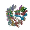 8vrdC  8vrkC 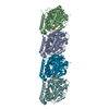 8vt7C M: map data used to model this data C: citing same article ( |
|---|---|
| Similar structure data |


-Links
 PDBj
PDBj


- Assembly
| Deposited unit | 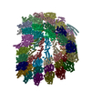
|
|---|---|
| 1 |
|
-Components
-Protein , 8 types, 53 molecules 1OPQRSTUVWXYZ2opqrstuvwxyz36L7...
| #1: Protein | Mass: 51019.297 Da / Num. of mol.: 13 / Mutation: E254D Source method: isolated from a genetically manipulated source Source: (gene. exp.) Homo sapiens (human) / Gene: TUBA1B / Production host:  Trichoplusia ni (cabbage looper) Trichoplusia ni (cabbage looper)References: UniProt: P68363, Hydrolases; Acting on acid anhydrides; Acting on GTP to facilitate cellular and subcellular movement #2: Protein | Mass: 51276.367 Da / Num. of mol.: 13 Source method: isolated from a genetically manipulated source Source: (gene. exp.) Homo sapiens (human) / Gene: TUBB3, TUBB4 / Production host: Trichoplusia ni (cabbage looper) / References: UniProt: Q13509#3: Protein | Mass: 199732.516 Da / Num. of mol.: 3 Source method: isolated from a genetically manipulated source Source: (gene. exp.) Homo sapiens (human) / Gene: TUBGCP6 / Production host: Trichoplusia ni (cabbage looper) / References: UniProt: B2RWN4#5: Protein | Mass: 8485.724 Da / Num. of mol.: 2 Source method: isolated from a genetically manipulated source Source: (gene. exp.) Homo sapiens (human) / Gene: MZT1, C13orf37, MOZART1 / Production host: Trichoplusia ni (cabbage looper) / References: UniProt: Q08AG7#6: Protein | | Mass: 41782.660 Da / Num. of mol.: 1 Source method: isolated from a genetically manipulated source Source: (gene. exp.) Homo sapiens (human) / Gene: ACTB / Production host: Trichoplusia ni (cabbage looper) / References: UniProt: P60709#7: Protein | Mass: 105581.500 Da / Num. of mol.: 5 Source method: isolated from a genetically manipulated source Source: (gene. exp.) Homo sapiens (human) / Gene: TUBGCP2, GCP2 / Production host: Trichoplusia ni (cabbage looper) / References: UniProt: Q9BSJ2#8: Protein | Mass: 76108.898 Da / Num. of mol.: 2 Source method: isolated from a genetically manipulated source Source: (gene. exp.) Homo sapiens (human) / Gene: TUBGCP4, 76P, GCP4 / Production host: Trichoplusia ni (cabbage looper) / References: UniProt: Q9UGJ1#10: Protein | Mass: 52022.617 Da / Num. of mol.: 14 Source method: isolated from a genetically manipulated source Source: (gene. exp.) Homo sapiens (human) / Gene: TUBG1, TUBG / Production host: Trichoplusia ni (cabbage looper) / References: UniProt: P23258 |
|---|
-Gamma-tubulin complex component ... , 2 types, 7 molecules 5BDFHNJ
| #4: Protein | Mass: 103710.102 Da / Num. of mol.: 6 Source method: isolated from a genetically manipulated source Source: (gene. exp.) Homo sapiens (human) / Gene: TUBGCP3, GCP3 / Production host: Trichoplusia ni (cabbage looper) / References: UniProt: Q96CW5#9: Protein | | Mass: 118367.406 Da / Num. of mol.: 1 Source method: isolated from a genetically manipulated source Source: (gene. exp.) Homo sapiens (human) / Gene: TUBGCP5, GCP5, KIAA1899 / Production host: Trichoplusia ni (cabbage looper) / References: UniProt: Q96RT8 |
|---|
-Experimental details
-Experiment
| Experiment | Method: ELECTRON MICROSCOPY |
|---|---|
| EM experiment | Aggregation state: PARTICLE / 3D reconstruction method: single particle reconstruction |
- Sample preparation
| Component | Name: Gamma tubulin ring complex bound to microtubule minus end Type: COMPLEX / Entity ID: all / Source: RECOMBINANT |
|---|---|
| Source (natural) | Organism: Homo sapiens (human) |
| Source (recombinant) | Organism: Trichoplusia ni (cabbage looper) |
| Buffer solution | pH: 6.8 |
| Specimen | Embedding applied: NO / Shadowing applied: NO / Staining applied: NO / Vitrification applied: YES |
| Vitrification | Cryogen name: ETHANE / Humidity: 100 % |
- Electron microscopy imaging
| Experimental equipment |  Model: Titan Krios / Image courtesy: FEI Company |
|---|---|
| Microscopy | Model: FEI TITAN KRIOS |
| Electron gun | Electron source:  FIELD EMISSION GUN / Accelerating voltage: 300 kV / Illumination mode: FLOOD BEAM FIELD EMISSION GUN / Accelerating voltage: 300 kV / Illumination mode: FLOOD BEAM |
| Electron lens | Mode: BRIGHT FIELD / Nominal defocus max: 3500 nm / Nominal defocus min: 1500 nm |
| Image recording | Electron dose: 60 e/Å2 / Film or detector model: GATAN K3 (6k x 4k) |
- Processing
| CTF correction | Type: PHASE FLIPPING AND AMPLITUDE CORRECTION |
|---|---|
| 3D reconstruction | Resolution: 7.7 Å / Resolution method: FSC 0.143 CUT-OFF / Num. of particles: 32375 / Symmetry type: POINT |
| Atomic model building | Protocol: RIGID BODY FIT |
