 Movie
Movie Controller

[English] 日本語
 Yorodumi
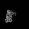
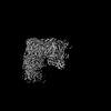
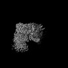
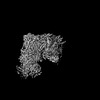
- PDB-8ugl: High resolution in-situ structure of complex IV in respiratory su... -
+ Open data

- Basic information
| Entry | Database: PDB / ID: 8ugl | ||||||
|---|---|---|---|---|---|---|---|
| Title | High resolution in-situ structure of complex IV in respiratory supercomplex | ||||||
 Components Components | (Cytochrome c oxidase subunit ...) x 14 | ||||||
 Keywords Keywords | ELECTRON TRANSPORT / in-situ cryo-EM structure / mammalian / mitochondria / respiratory supercomplex / proton pumping / membrane protein | ||||||
| Function / homology |  Function and homology information Function and homology informationComplex IV assembly / Cytoprotection by HMOX1 / TP53 Regulates Metabolic Genes / respiratory chain complex IV assembly / mitochondrial respirasome assembly / Respiratory electron transport / Mitochondrial translation termination / Mitochondrial protein degradation / respiratory chain complex IV / respiratory chain complex ...Complex IV assembly / Cytoprotection by HMOX1 / TP53 Regulates Metabolic Genes / respiratory chain complex IV assembly / mitochondrial respirasome assembly / Respiratory electron transport / Mitochondrial translation termination / Mitochondrial protein degradation / respiratory chain complex IV / respiratory chain complex / cytochrome-c oxidase / oxidative phosphorylation / mitochondrial electron transport, cytochrome c to oxygen / cytochrome-c oxidase activity / positive regulation of vasoconstriction / ATP synthesis coupled electron transport / enzyme regulator activity / aerobic respiration / respiratory electron transport chain / central nervous system development / mitochondrial membrane / oxidoreductase activity / mitochondrial inner membrane / copper ion binding / heme binding / mitochondrion / metal ion binding Similarity search - Function | ||||||
| Biological species |  | ||||||
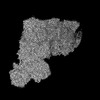
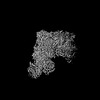
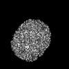
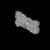
| Method | ELECTRON MICROSCOPY / single particle reconstruction / cryo EM / Resolution: 3 Å | ||||||
 Authors Authors | Zheng, W. / Zhu, J. / Zhang, K. | ||||||
| Funding support |  United States, 1items United States, 1items
| ||||||
 Citation Citation | Journal: Nature / Year: 2024 Title: High-resolution in situ structures of mammalian respiratory supercomplexes. Authors: Wan Zheng / Pengxin Chai / Jiapeng Zhu / Kai Zhang /  Abstract: Mitochondria play a pivotal part in ATP energy production through oxidative phosphorylation, which occurs within the inner membrane through a series of respiratory complexes. Despite extensive in ...Mitochondria play a pivotal part in ATP energy production through oxidative phosphorylation, which occurs within the inner membrane through a series of respiratory complexes. Despite extensive in vitro structural studies, determining the atomic details of their molecular mechanisms in physiological states remains a major challenge, primarily because of loss of the native environment during purification. Here we directly image porcine mitochondria using an in situ cryo-electron microscopy approach. This enables us to determine the structures of various high-order assemblies of respiratory supercomplexes in their native states. We identify four main supercomplex organizations: IIIIIV, IIIIIV, IIIIIV and IIIIIV, which potentially expand into higher-order arrays on the inner membranes. These diverse supercomplexes are largely formed by 'protein-lipids-protein' interactions, which in turn have a substantial impact on the local geometry of the surrounding membranes. Our in situ structures also capture numerous reactive intermediates within these respiratory supercomplexes, shedding light on the dynamic processes of the ubiquinone/ubiquinol exchange mechanism in complex I and the Q-cycle in complex III. Structural comparison of supercomplexes from mitochondria treated under different conditions indicates a possible correlation between conformational states of complexes I and III, probably in response to environmental changes. By preserving the native membrane environment, our approach enables structural studies of mitochondrial respiratory supercomplexes in reaction at high resolution across multiple scales, from atomic-level details to the broader subcellular context. | ||||||
| History |
|
- Structure visualization
| Structure viewer | Molecule: MolmilJmol/JSmol |
|---|
- Downloads & links
-Download
| PDBx/mmCIF format | 8ugl.cif.gz | 442.3 KB | Display | PDBx/mmCIF format |
|---|---|---|---|---|
| PDB format | pdb8ugl.ent.gz | Display | PDB format | |
| PDBx/mmJSON format | 8ugl.json.gz | Tree view | PDBx/mmJSON format | |
| Others |  Other downloads Other downloads |
-Validation report
| Arichive directory | https://data.pdbj.org/pub/pdb/validation_reports/ug/8uglftp://data.pdbj.org/pub/pdb/validation_reports/ug/8ugl | HTTPS FTP |
|---|
-Related structure data
| Related structure data | 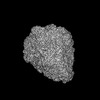 42229MC  8ud1C  8ueoC  8uepC  8ueqC  8uerC  8uesC  8uetC  8ueuC  8uevC  8uewC  8uexC  8ueyC  8uezC  8ugdC  8ugeC  8ugfC  8uggC  8ughC  8ugiC  8ugjC  8ugkC  8ugnC 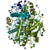 8ugpC  8ugrC M: map data used to model this data C: citing same article ( |
|---|---|
| Similar structure data |


-Links
 PDBj
PDBj


- Assembly
| Deposited unit | 
|
|---|---|
| 1 |
|
-Components
-Cytochrome c oxidase subunit ... , 14 types, 14 molecules 4A4B4C4D4E4F4G4H4I4J4K4L4M4N
| #1: Protein | Mass: 56992.711 Da / Num. of mol.: 1 / Source method: isolated from a natural source / Source: (natural) |
|---|---|
| #2: Protein | Mass: 26288.627 Da / Num. of mol.: 1 / Source method: isolated from a natural source / Source: (natural) |
| #3: Protein | Mass: 29753.361 Da / Num. of mol.: 1 / Source method: isolated from a natural source / Source: (natural) |
| #4: Protein | Mass: 19670.664 Da / Num. of mol.: 1 / Source method: isolated from a natural source / Source: (natural) |
| #5: Protein | Mass: 16771.195 Da / Num. of mol.: 1 / Source method: isolated from a natural source / Source: (natural) |
| #6: Protein | Mass: 13801.783 Da / Num. of mol.: 1 / Source method: isolated from a natural source / Source: (natural) |
| #7: Protein | Mass: 10876.370 Da / Num. of mol.: 1 / Source method: isolated from a natural source / Source: (natural) |
| #8: Protein | Mass: 10204.456 Da / Num. of mol.: 1 / Source method: isolated from a natural source / Source: (natural) |
| #9: Protein | Mass: 8617.122 Da / Num. of mol.: 1 / Source method: isolated from a natural source / Source: (natural) |
| #10: Protein | Mass: 9028.479 Da / Num. of mol.: 1 / Source method: isolated from a natural source / Source: (natural) |
| #11: Protein | Mass: 9169.476 Da / Num. of mol.: 1 / Source method: isolated from a natural source / Source: (natural) |
| #12: Protein | Mass: 7358.661 Da / Num. of mol.: 1 / Source method: isolated from a natural source / Source: (natural) |
| #13: Protein | Mass: 7615.939 Da / Num. of mol.: 1 / Source method: isolated from a natural source / Source: (natural) |
| #14: Protein | Mass: 9322.771 Da / Num. of mol.: 1 / Source method: isolated from a natural source / Source: (natural) |
-Non-polymers , 12 types, 833 molecules 
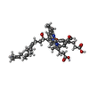


| #15: Chemical | ChemComp-PGV / (  Mass: 749.007 Da / Num. of mol.: 15 / Source method: obtained synthetically / Formula: C40H77O10P / Comment: phospholipid*YM Mass: 749.007 Da / Num. of mol.: 15 / Source method: obtained synthetically / Formula: C40H77O10P / Comment: phospholipid*YM#16: Chemical |  Mass: 852.837 Da / Num. of mol.: 2 / Source method: obtained synthetically / Formula: C49H56FeN4O6 / Feature type: SUBJECT OF INVESTIGATION Mass: 852.837 Da / Num. of mol.: 2 / Source method: obtained synthetically / Formula: C49H56FeN4O6 / Feature type: SUBJECT OF INVESTIGATION#17: Chemical | ChemComp-CU / |  Mass: 63.546 Da / Num. of mol.: 1 / Source method: obtained synthetically / Formula: Cu Mass: 63.546 Da / Num. of mol.: 1 / Source method: obtained synthetically / Formula: Cu#18: Chemical | ChemComp-MG / |  Mass: 24.305 Da / Num. of mol.: 1 / Source method: obtained synthetically / Formula: Mg Mass: 24.305 Da / Num. of mol.: 1 / Source method: obtained synthetically / Formula: Mg#19: Chemical | ChemComp-NA / |  Mass: 22.990 Da / Num. of mol.: 1 / Source method: obtained synthetically / Formula: Na Mass: 22.990 Da / Num. of mol.: 1 / Source method: obtained synthetically / Formula: Na#20: Chemical |  Mass: 1464.043 Da / Num. of mol.: 3 / Source method: obtained synthetically / Formula: C81H156O17P2 / Comment: phospholipid*YM Mass: 1464.043 Da / Num. of mol.: 3 / Source method: obtained synthetically / Formula: C81H156O17P2 / Comment: phospholipid*YM#21: Chemical | ChemComp-CUA / |  Mass: 127.092 Da / Num. of mol.: 1 / Source method: obtained synthetically / Formula: Cu2 Mass: 127.092 Da / Num. of mol.: 1 / Source method: obtained synthetically / Formula: Cu2#22: Chemical | ChemComp-PSC / ( |  Mass: 759.068 Da / Num. of mol.: 1 / Source method: obtained synthetically / Formula: C42H81NO8P / Comment: phospholipid*YM Mass: 759.068 Da / Num. of mol.: 1 / Source method: obtained synthetically / Formula: C42H81NO8P / Comment: phospholipid*YM#23: Chemical | ChemComp-ZN / |  Mass: 65.409 Da / Num. of mol.: 1 / Source method: obtained synthetically / Formula: Zn Mass: 65.409 Da / Num. of mol.: 1 / Source method: obtained synthetically / Formula: Zn#24: Chemical |  Mass: 768.055 Da / Num. of mol.: 2 / Source method: obtained synthetically / Formula: C43H78NO8P / Comment: phospholipid*YM Mass: 768.055 Da / Num. of mol.: 2 / Source method: obtained synthetically / Formula: C43H78NO8P / Comment: phospholipid*YM#25: Chemical | ChemComp-PO4 / |  Mass: 94.971 Da / Num. of mol.: 1 / Source method: obtained synthetically / Formula: PO4 Mass: 94.971 Da / Num. of mol.: 1 / Source method: obtained synthetically / Formula: PO4#26: Water | ChemComp-HOH / | Mass: 18.015 Da / Num. of mol.: 804 / Source method: isolated from a natural source / Formula: H2O |
|---|
-Details
| Has ligand of interest | Y |
|---|---|
| Has protein modification | Y |
-Experimental details
-Experiment
| Experiment | Method: ELECTRON MICROSCOPY |
|---|---|
| EM experiment | Aggregation state: PARTICLE / 3D reconstruction method: single particle reconstruction |
- Sample preparation
| Component | Name: In-situ cryo-EM structure of respiratory supercomplex by directly imaging mitochondria Type: ORGANELLE OR CELLULAR COMPONENT / Entity ID: #1-#14 / Source: NATURAL |
|---|---|
| Source (natural) | Organism: |
| Buffer solution | pH: 7 |
| Specimen | Embedding applied: NO / Shadowing applied: NO / Staining applied: NO / Vitrification applied: YES |
| Vitrification | Cryogen name: ETHANE |
- Electron microscopy imaging
| Experimental equipment |  Model: Titan Krios / Image courtesy: FEI Company |
|---|---|
| Microscopy | Model: FEI TITAN KRIOS |
| Electron gun | Electron source:  FIELD EMISSION GUN / Accelerating voltage: 300 kV / Illumination mode: FLOOD BEAM FIELD EMISSION GUN / Accelerating voltage: 300 kV / Illumination mode: FLOOD BEAM |
| Electron lens | Mode: BRIGHT FIELD / Nominal defocus max: 3000 nm / Nominal defocus min: 1300 nm / Cs: 2.7 mm |
| Image recording | Electron dose: 50 e/Å2 / Film or detector model: GATAN K3 BIOQUANTUM (6k x 4k) |
- Processing
| EM software | Name: PHENIX / Version: 1.20.1_4487: / Category: model refinement | ||||||||||||||||||||||||
|---|---|---|---|---|---|---|---|---|---|---|---|---|---|---|---|---|---|---|---|---|---|---|---|---|---|
| CTF correction | Type: PHASE FLIPPING AND AMPLITUDE CORRECTION | ||||||||||||||||||||||||
| 3D reconstruction | Resolution: 3 Å / Resolution method: FSC 0.143 CUT-OFF / Num. of particles: 56000 / Symmetry type: POINT | ||||||||||||||||||||||||
| Refine LS restraints |
|
