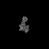
 Movie
Movie Controller


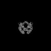
+ Open data

- Basic information
| Entry | Database: PDB / ID: 8u8b | ||||||
|---|---|---|---|---|---|---|---|
| Title | Cryo-EM structure of LRRK2 bound to type II inhibitor rebastinib | ||||||
 Components Components | Leucine-rich repeat serine/threonine-protein kinase 2 | ||||||
 Keywords Keywords | HYDROLASE/HYDROLASE INHIBITOR / Cryo-EM / Parkinson's disease / Kinase / LRRK2 / type II inhibitor / HYDROLASE / HYDROLASE-HYDROLASE INHIBITOR complex | ||||||
| Function / homology |  Function and homology information Function and homology informationcaveola neck / : / beta-catenin destruction complex binding / regulation of branching morphogenesis of a nerve / Wnt signalosome assembly / negative regulation of motile cilium assembly / regulation of kidney size / regulation of cell projection organization / tangential migration from the subventricular zone to the olfactory bulb / GTP-dependent protein kinase activity ...caveola neck / : / beta-catenin destruction complex binding / regulation of branching morphogenesis of a nerve / Wnt signalosome assembly / negative regulation of motile cilium assembly / regulation of kidney size / regulation of cell projection organization / tangential migration from the subventricular zone to the olfactory bulb / GTP-dependent protein kinase activity / regulation of SNARE complex assembly / regulation of neuroblast proliferation / regulation of ER to Golgi vesicle-mediated transport / protein localization to endoplasmic reticulum exit site / peroxidase inhibitor activity / negative regulation of late endosome to lysosome transport / regulation of mitochondrial depolarization / : / positive regulation of dopamine receptor signaling pathway / amphisome / regulation of synaptic vesicle transport / : / regulation of CAMKK-AMPK signaling cascade / co-receptor binding / negative regulation of GTPase activity / regulation of dopamine receptor signaling pathway / positive regulation of microglial cell activation / regulation of retrograde transport, endosome to Golgi / regulation of neuron maturation / cellular response to curcumin / olfactory bulb development / cytoplasmic side of mitochondrial outer membrane / positive regulation of synaptic vesicle endocytosis / JUN kinase kinase kinase activity / negative regulation of autophagosome assembly / multivesicular body, internal vesicle / regulation of cAMP/PKA signal transduction / negative regulation of excitatory postsynaptic potential / striatum development / neuron projection arborization / mitochondrion localization / regulation of dendritic spine morphogenesis / protein localization to mitochondrion / cellular response to dopamine / positive regulation of mitochondrial outer membrane permeabilization involved in apoptotic signaling pathway / endoplasmic reticulum organization / negative regulation of protein processing / positive regulation of protein autoubiquitination / Wnt signalosome / positive regulation of programmed cell death / GTP metabolic process / regulation of canonical Wnt signaling pathway / syntaxin-1 binding / regulation of reactive oxygen species metabolic process / Golgi-associated vesicle / PTK6 promotes HIF1A stabilization / lysosome organization / clathrin binding / negative regulation of macroautophagy / regulation of mitochondrial fission / Lewy body / regulation of synaptic vesicle exocytosis / regulation of locomotion / intracellular distribution of mitochondria / Golgi organization / neuromuscular junction development / protein kinase A binding / microvillus / exploration behavior / autolysosome / locomotory exploration behavior / endoplasmic reticulum exit site / negative regulation of Notch signaling pathway / MAP kinase kinase kinase activity / regulation of synaptic vesicle endocytosis / canonical Wnt signaling pathway / negative regulation of endoplasmic reticulum stress-induced intrinsic apoptotic signaling pathway / Rho protein signal transduction / regulation of synaptic transmission, glutamatergic / presynaptic cytosol / JNK cascade / phagocytic vesicle / cellular response to manganese ion / neuron projection morphogenesis / positive regulation of autophagy / dendrite cytoplasm / cellular response to starvation / positive regulation of protein ubiquitination / GTPase activator activity / determination of adult lifespan / regulation of autophagy / SNARE binding / cellular response to reactive oxygen species / mitochondrion organization / trans-Golgi network / calcium-mediated signaling / regulation of protein stability / regulation of membrane potential / tubulin binding / excitatory postsynaptic potential Similarity search - Function | ||||||
| Biological species |  Homo sapiens (human) Homo sapiens (human) | ||||||
| Method | ELECTRON MICROSCOPY / single particle reconstruction / cryo EM / Resolution: 3.7 Å | ||||||
 Authors Authors | Zhu, H. / Sun, J. | ||||||
| Funding support |  United States, 1items United States, 1items
| ||||||
 Citation Citation | Journal: Cell Discov / Year: 2024 Title: Pharmacology of LRRK2 with type I and II kinase inhibitors revealed by cryo-EM. Authors: Hanwen Zhu / Patricia Hixson / Wen Ma / Ji Sun / Abstract: LRRK2 is one of the most promising drug targets for Parkinson's disease. Though type I kinase inhibitors of LRRK2 are under clinical trials, alternative strategies like type II inhibitors are being ...LRRK2 is one of the most promising drug targets for Parkinson's disease. Though type I kinase inhibitors of LRRK2 are under clinical trials, alternative strategies like type II inhibitors are being actively pursued due to the potential undesired effects of type I inhibitors. Currently, a robust method for LRRK2-inhibitor structure determination to guide structure-based drug discovery is lacking, and inhibition mechanisms of available compounds are also unclear. Here we present near-atomic-resolution structures of LRRK2 with type I (LRRK2-IN-1 and GNE-7915) and type II (rebastinib, ponatinib, and GZD-824) inhibitors, uncovering the structural basis of LRRK2 inhibition and conformational plasticity of the kinase domain with molecular dynamics (MD) simulations. Type I and II inhibitors bind to LRRK2 in active-like and inactive conformations, so LRRK2-inhibitor complexes further reveal general structural features associated with LRRK2 activation. Our study provides atomic details of LRRK2-inhibitor interactions and a framework for understanding LRRK2 activation and for rational drug design. | ||||||
| History |
|
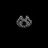
- Structure visualization
| Structure viewer | Molecule: MolmilJmol/JSmol |
|---|
- Downloads & links
-Download
| PDBx/mmCIF format | 8u8b.cif.gz | 597.3 KB | Display | PDBx/mmCIF format |
|---|---|---|---|---|
| PDB format | pdb8u8b.ent.gz | 454.7 KB | Display | PDB format |
| PDBx/mmJSON format | 8u8b.json.gz | Tree view | PDBx/mmJSON format | |
| Others |  Other downloads Other downloads |
-Validation report
| Arichive directory | https://data.pdbj.org/pub/pdb/validation_reports/u8/8u8bftp://data.pdbj.org/pub/pdb/validation_reports/u8/8u8b | HTTPS FTP |
|---|
-Related structure data
| Related structure data |  42020MC 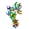 8fo7C 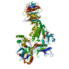 8u7hC  8u7lC  8u8aC M: map data used to model this data C: citing same article ( |
|---|---|
| Similar structure data |
-Links
 PDBj
PDBj


- Assembly
| Deposited unit | 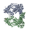
|
|---|---|
| 1 |
|
-Components
| #1: Protein | Mass: 286427.656 Da / Num. of mol.: 2 Source method: isolated from a genetically manipulated source Source: (gene. exp.) Homo sapiens (human) / Gene: LRRK2, PARK8 / Production host: Homo sapiens (human)References: UniProt: Q5S007, non-specific serine/threonine protein kinase, Hydrolases; Acting on acid anhydrides; Acting on GTP to facilitate cellular and subcellular movement #2: Chemical |   Type: RNA linking / Mass: 443.201 Da / Num. of mol.: 2 / Source method: obtained synthetically / Formula: C10H15N5O11P2 / Comment: GDP, energy-carrying molecule*YM Type: RNA linking / Mass: 443.201 Da / Num. of mol.: 2 / Source method: obtained synthetically / Formula: C10H15N5O11P2 / Comment: GDP, energy-carrying molecule*YM#3: Chemical | 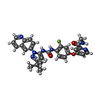  Mass: 553.587 Da / Num. of mol.: 2 / Source method: obtained synthetically / Formula: C30H28FN7O3 / Feature type: SUBJECT OF INVESTIGATION Mass: 553.587 Da / Num. of mol.: 2 / Source method: obtained synthetically / Formula: C30H28FN7O3 / Feature type: SUBJECT OF INVESTIGATIONHas ligand of interest | Y | |
|---|
-Experimental details
-Experiment
| Experiment | Method: ELECTRON MICROSCOPY |
|---|---|
| EM experiment | Aggregation state: PARTICLE / 3D reconstruction method: single particle reconstruction |
- Sample preparation
| Component | Name: LRRK2-rebastinib / Type: COMPLEX / Entity ID: #1 / Source: RECOMBINANT |
|---|---|
| Molecular weight | Value: 286 kDa/nm / Experimental value: YES |
| Source (natural) | Organism: Homo sapiens (human) |
| Source (recombinant) | Organism: Homo sapiens (human) |
| Buffer solution | pH: 8 |
| Specimen | Embedding applied: NO / Shadowing applied: NO / Staining applied: NO / Vitrification applied: YES |
| Vitrification | Cryogen name: ETHANE |
- Electron microscopy imaging
| Experimental equipment |  Model: Titan Krios / Image courtesy: FEI Company |
|---|---|
| Microscopy | Model: FEI TITAN KRIOS |
| Electron gun | Electron source:  FIELD EMISSION GUN / Accelerating voltage: 300 kV / Illumination mode: OTHER FIELD EMISSION GUN / Accelerating voltage: 300 kV / Illumination mode: OTHER |
| Electron lens | Mode: BRIGHT FIELD / Nominal defocus max: 1800 nm / Nominal defocus min: 600 nm |
| Image recording | Electron dose: 67.53 e/Å2 / Film or detector model: GATAN K3 (6k x 4k) |
- Processing
| EM software |
| ||||||||||||
|---|---|---|---|---|---|---|---|---|---|---|---|---|---|
| CTF correction | Type: NONE | ||||||||||||
| 3D reconstruction | Resolution: 3.7 Å / Resolution method: FSC 0.143 CUT-OFF / Num. of particles: 75005 / Symmetry type: POINT |
