 Movie
Movie Controller


+ Open data

- Basic information
| Entry | Database: PDB / ID: 8u4j | ||||||||||||
|---|---|---|---|---|---|---|---|---|---|---|---|---|---|
| Title | Structure of the HER4/BTC Homodimer Extracellular Domain | ||||||||||||
 Components Components |
| ||||||||||||
 Keywords Keywords | MEMBRANE PROTEIN / TRANSFERASE / Receptor Tyrosine Kinase | ||||||||||||
| Function / homology |  Function and homology information Function and homology informationERBB4 signaling pathway / olfactory bulb interneuron differentiation / ERBB4-ERBB4 signaling pathway / central nervous system morphogenesis / neuregulin receptor activity / cardiac muscle tissue regeneration / mammary gland epithelial cell differentiation / mammary gland alveolus development / ERBB2-ERBB4 signaling pathway / mitochondrial fragmentation involved in apoptotic process ...ERBB4 signaling pathway / olfactory bulb interneuron differentiation / ERBB4-ERBB4 signaling pathway / central nervous system morphogenesis / neuregulin receptor activity / cardiac muscle tissue regeneration / mammary gland epithelial cell differentiation / mammary gland alveolus development / ERBB2-ERBB4 signaling pathway / mitochondrial fragmentation involved in apoptotic process / transmembrane receptor protein tyrosine kinase activator activity / embryonic pattern specification / PI3K events in ERBB4 signaling / neural crest cell migration / GABA receptor binding / Inhibition of Signaling by Overexpressed EGFR / EGFR interacts with phospholipase C-gamma / epidermal growth factor receptor activity / epidermal growth factor receptor binding / ERBB2-EGFR signaling pathway / ERBB2 Activates PTK6 Signaling / positive regulation of tyrosine phosphorylation of STAT protein / Signaling by EGFR / neurotransmitter receptor localization to postsynaptic specialization membrane / ERBB2 Regulates Cell Motility / positive regulation of cell division / Signaling by ERBB4 / PI3K events in ERBB2 signaling / Long-term potentiation / Estrogen-dependent nuclear events downstream of ESR-membrane signaling / GAB1 signalosome / SHC1 events in ERBB4 signaling / positive regulation of cardiac muscle cell proliferation / Nuclear signaling by ERBB4 / lactation / cell surface receptor signaling pathway via JAK-STAT / Signaling by ERBB2 / positive regulation of mitotic nuclear division / transmembrane receptor protein tyrosine kinase activity / synapse assembly / peptidyl-tyrosine phosphorylation / regulation of cell migration / GRB2 events in EGFR signaling / SHC1 events in EGFR signaling / positive regulation of epithelial cell proliferation / GRB2 events in ERBB2 signaling / SHC1 events in ERBB2 signaling / cell surface receptor protein tyrosine kinase signaling pathway / basal plasma membrane / Downregulation of ERBB4 signaling / positive regulation of receptor signaling pathway via JAK-STAT / growth factor activity / neuromuscular junction / clathrin-coated endocytic vesicle membrane / Signaling by ERBB2 TMD/JMD mutants / EGFR downregulation / receptor protein-tyrosine kinase / Signaling by ERBB2 KD Mutants / positive regulation of protein phosphorylation / postsynaptic density membrane / GABA-ergic synapse / epidermal growth factor receptor signaling pathway / Downregulation of ERBB2 signaling / neuron differentiation / Constitutive Signaling by Aberrant PI3K in Cancer / heart development / PIP3 activates AKT signaling / protein autophosphorylation / cell migration / Cargo recognition for clathrin-mediated endocytosis / nervous system development / Clathrin-mediated endocytosis / PI5P, PP2A and IER3 Regulate PI3K/AKT Signaling / RAF/MAP kinase cascade / protein tyrosine kinase activity / presynaptic membrane / Estrogen-dependent gene expression / basolateral plasma membrane / positive regulation of MAPK cascade / positive regulation of ERK1 and ERK2 cascade / signaling receptor complex / postsynaptic membrane / Extra-nuclear estrogen signaling / transcription cis-regulatory region binding / mitochondrial matrix / negative regulation of cell population proliferation / receptor ligand activity / positive regulation of cell population proliferation / negative regulation of apoptotic process / positive regulation of DNA-templated transcription / glutamatergic synapse / signal transduction / protein homodimerization activity / mitochondrion / DNA-templated transcription / : / extracellular region / nucleoplasm / ATP binding / nucleus Similarity search - Function | ||||||||||||
| Biological species |  Homo sapiens (human) Homo sapiens (human) | ||||||||||||
| Method | ELECTRON MICROSCOPY / single particle reconstruction / cryo EM / Resolution: 3.7 Å | ||||||||||||
 Authors Authors | Trenker, R. / Diwanji, D. / Bingham, T. / Verba, K.A. / Jura, N. | ||||||||||||
| Funding support |  Germany, Germany,  United States, 3items United States, 3items
| ||||||||||||
 Citation Citation | Journal: Elife / Year: 2024 Title: Structural dynamics of the active HER4 and HER2/HER4 complexes is finely tuned by different growth factors and glycosylation. Authors: Raphael Trenker / Devan Diwanji / Tanner Bingham / Kliment A Verba / Natalia Jura / Abstract: Human Epidermal growth factor Receptor 4 (HER4 or ERBB4) carries out essential functions in the development and maintenance of the cardiovascular and nervous systems. HER4 activation is regulated by ...Human Epidermal growth factor Receptor 4 (HER4 or ERBB4) carries out essential functions in the development and maintenance of the cardiovascular and nervous systems. HER4 activation is regulated by a diverse group of extracellular ligands including the neuregulin (NRG) family and betacellulin (BTC), which promote HER4 homodimerization or heterodimerization with other HER receptors. Important cardiovascular functions of HER4 are exerted via heterodimerization with its close homolog and orphan receptor, HER2. To date structural insights into ligand-mediated HER4 activation have been limited to crystallographic studies of HER4 ectodomain homodimers in complex with NRG1β. Here, we report cryo-EM structures of near full-length HER2/HER4 heterodimers and full-length HER4 homodimers bound to NRG1β and BTC. We show that the structures of the heterodimers bound to either ligand are nearly identical and that in both cases the HER2/HER4 heterodimer interface is less dynamic than those observed in structures of HER2/EGFR and HER2/HER3 heterodimers. In contrast, structures of full-length HER4 homodimers bound to NRG1β and BTC display more large-scale dynamics mirroring states previously reported for EGFR homodimers. Our structures also reveal the presence of multiple glycan modifications within HER4 ectodomains, modeled for the first time in HER receptors, that distinctively contribute to the stabilization of HER4 homodimer interfaces over those of HER2/HER4 heterodimers. | ||||||||||||
| History |
|
- Structure visualization
| Structure viewer | Molecule: MolmilJmol/JSmol |
|---|
- Downloads & links
-Download
| PDBx/mmCIF format | 8u4j.cif.gz | 481.3 KB | Display | PDBx/mmCIF format |
|---|---|---|---|---|
| PDB format | pdb8u4j.ent.gz | 414.4 KB | Display | PDB format |
| PDBx/mmJSON format | 8u4j.json.gz | Tree view | PDBx/mmJSON format | |
| Others |  Other downloads Other downloads |
-Validation report
| Arichive directory | https://data.pdbj.org/pub/pdb/validation_reports/u4/8u4jftp://data.pdbj.org/pub/pdb/validation_reports/u4/8u4j | HTTPS FTP |
|---|
-Related structure data
| Related structure data |  41884MC  8u4iC  8u4kC 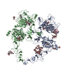 8u4lC M: map data used to model this data C: citing same article ( |
|---|---|
| Similar structure data |


-Links
 PDBj
PDBj










- Assembly
| Deposited unit | 
|
|---|---|
| 1 |
|
-Components
-Protein / Protein/peptide , 2 types, 4 molecules ABCD
| #1: Protein | Mass: 68072.820 Da / Num. of mol.: 2 / Fragment: intracellular domain (UNP residues 26-635) Source method: isolated from a genetically manipulated source Source: (gene. exp.) Homo sapiens (human) / Gene: ERBB4, HER4 / Plasmid: pCDNA4 / Cell line (production host): EXPI293F / Production host: Homo sapiens (human)References: UniProt: Q15303, receptor protein-tyrosine kinase #2: Protein/peptide | Mass: 5630.513 Da / Num. of mol.: 2 Source method: isolated from a genetically manipulated source Source: (gene. exp.) Homo sapiens (human) / Gene: BTC / Plasmid: pET32 / Production host:   Escherichia coli BL21(DE3) (bacteria) / Strain (production host): Origami B (DE3) / References: UniProt: P35070 Escherichia coli BL21(DE3) (bacteria) / Strain (production host): Origami B (DE3) / References: UniProt: P35070 |
|---|
-Sugars , 4 types, 16 molecules 
| #3: Polysaccharide | beta-D-mannopyranose-(1-4)-2-acetamido-2-deoxy-beta-D-glucopyranose-(1-4)-2-acetamido-2-deoxy-beta- ...beta-D-mannopyranose-(1-4)-2-acetamido-2-deoxy-beta-D-glucopyranose-(1-4)-2-acetamido-2-deoxy-beta-D-glucopyranose Source method: isolated from a genetically manipulated source #4: Polysaccharide | alpha-D-mannopyranose-(1-3)-[alpha-D-mannopyranose-(1-6)]beta-D-mannopyranose-(1-4)-2-acetamido-2- ...alpha-D-mannopyranose-(1-3)-[alpha-D-mannopyranose-(1-6)]beta-D-mannopyranose-(1-4)-2-acetamido-2-deoxy-beta-D-glucopyranose-(1-4)-2-acetamido-2-deoxy-beta-D-glucopyranose Source method: isolated from a genetically manipulated source #5: Polysaccharide | Source method: isolated from a genetically manipulated source #6: Sugar |  Type: D-saccharide, beta linking / Mass: 221.208 Da / Num. of mol.: 2 / Source method: obtained synthetically / Formula: C8H15NO6 Type: D-saccharide, beta linking / Mass: 221.208 Da / Num. of mol.: 2 / Source method: obtained synthetically / Formula: C8H15NO6 |
|---|
-Details
| Has ligand of interest | N |
|---|---|
| Has protein modification | Y |
-Experimental details
-Experiment
| Experiment | Method: ELECTRON MICROSCOPY |
|---|---|
| EM experiment | Aggregation state: PARTICLE / 3D reconstruction method: single particle reconstruction |
- Sample preparation
| Component | Name: HER4/BTC homodimer / Type: COMPLEX / Entity ID: #1-#2 / Source: MULTIPLE SOURCES | ||||||||||||||||||||
|---|---|---|---|---|---|---|---|---|---|---|---|---|---|---|---|---|---|---|---|---|---|
| Molecular weight | Value: 0.306694 MDa / Experimental value: NO | ||||||||||||||||||||
| Source (natural) | Organism: Homo sapiens (human) | ||||||||||||||||||||
| Source (recombinant) | Organism: Homo sapiens (human) / Strain: EXPI293F / Plasmid: pCDNA4 | ||||||||||||||||||||
| Buffer solution | pH: 7.4 | ||||||||||||||||||||
| Buffer component |
| ||||||||||||||||||||
| Specimen | Embedding applied: NO / Shadowing applied: NO / Staining applied: NO / Vitrification applied: YES | ||||||||||||||||||||
| Specimen support | Grid type: Quantifoil R1.2/1.3 | ||||||||||||||||||||
| Vitrification | Instrument: FEI VITROBOT MARK IV / Cryogen name: ETHANE / Humidity: 100 % / Chamber temperature: 293 K |
- Electron microscopy imaging
| Experimental equipment |  Model: Titan Krios / Image courtesy: FEI Company |
|---|---|
| Microscopy | Model: TFS KRIOS |
| Electron gun | Electron source:  FIELD EMISSION GUN / Accelerating voltage: 300 kV / Illumination mode: FLOOD BEAM FIELD EMISSION GUN / Accelerating voltage: 300 kV / Illumination mode: FLOOD BEAM |
| Electron lens | Mode: BRIGHT FIELD / Nominal defocus max: 2000 nm / Nominal defocus min: 900 nm |
| Image recording | Electron dose: 45.8 e/Å2 / Film or detector model: GATAN K3 (6k x 4k) / Num. of grids imaged: 1 |
- Processing
| EM software |
| ||||||||||||||||||||||||
|---|---|---|---|---|---|---|---|---|---|---|---|---|---|---|---|---|---|---|---|---|---|---|---|---|---|
| CTF correction | Type: PHASE FLIPPING AND AMPLITUDE CORRECTION | ||||||||||||||||||||||||
| 3D reconstruction | Resolution: 3.7 Å / Resolution method: FSC 0.143 CUT-OFF / Num. of particles: 274540 / Symmetry type: POINT | ||||||||||||||||||||||||
| Atomic model building | PDB-ID: 3U7U Accession code: 3U7U / Source name: PDB / Type: experimental model |

